. 2004 Jan;113(2):285-301.
doi: 10.1172/JCI11548.

Translational upregulation of folate receptors is mediated by homocysteine via RNA-heterogeneous nuclear ribonucleoprotein E1 interactions

Affiliations

Translational upregulation of folate receptors is mediated by homocysteine via RNA-heterogeneous nuclear ribonucleoprotein E1 interactions

Aśok Antony et al. J Clin Invest. 2004 Jan.

Abstract

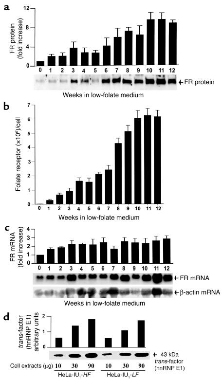
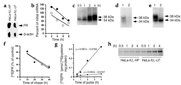
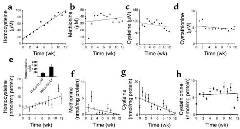
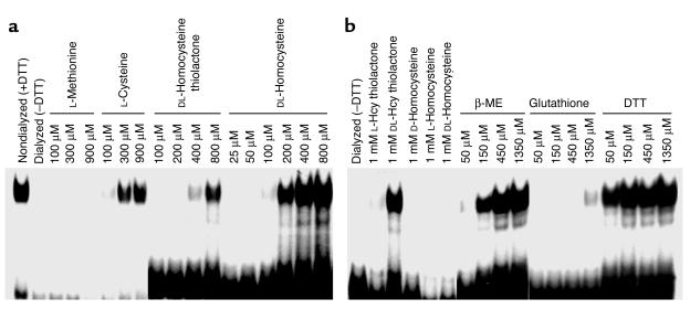
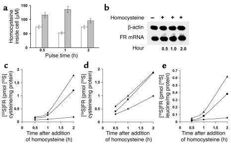
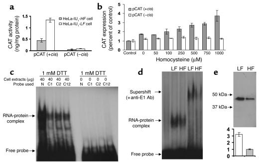
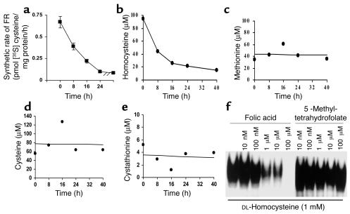
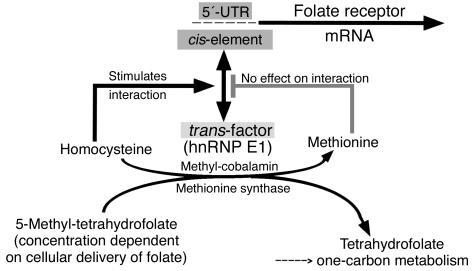
Cellular acquisition of folate is mediated by folate receptors (FRs) in many malignant and normal human cells. Although FRs are upregulated in folate deficiency and downregulated following folate repletion, the mechanistic basis for this relationship is unclear. Previously we demonstrated that interaction of an 18-base cis-element in the 5'-untranslated region of FR mRNA and a cystolic trans-factor (heterogeneous nuclear ribonucleoprotein E1 [hnRNP E1]) is critical for FR synthesis. However, the molecular mechanisms controlling this interaction, especially within the context of FR regulation and folate status, have remained obscure. Human cervical carcinoma cells exhibited progressively increasing upregulation of FRs after shifting of folate-replete cells to low-folate media, without a proportionate rise in FR mRNA or rise in hnRNP E1. Translational FR upregulation was accompanied by a progressive accumulation of the metabolite homocysteine within cultured cells, which stimulated interaction of the FR mRNA cis-element and hnRNP E1 as well as FR biosynthesis in a dose-dependent manner. Abrupt reversal of folate deficiency also led to a rapid parallel reduction in homocysteine and FR biosynthesis to levels observed in folate-replete cells. Collectively, these results suggest that homocysteine is the key modulator of translational upregulation of FRs and establishes the linkage between perturbed folate metabolism and coordinated upregulation of FRs.


Figures

Figure 1
Figure 1
Analysis of FR protein, FR mRNA, and hnRNP E1 in HeLa-IU1-HF cells propagated in low-folate media over 12 weeks. (a) Western blot analysis of the expression of FR protein as a function of time after shift of HeLa-IU1-HF cells to low-folate media. Each data point (fold increase of FR protein as a function of time) represents the mean ± SD of four independent evaluations. The Western blot data shown represent one of the four studies. (b) Determination of the cell surface receptor number by [3H]folate-binding to intact cells and Scatchard analysis as a function of time after shifting of HeLa-IU1-HF cells to low-folate media. Each data point represents the mean ± SD of four independent evaluations. (c) Northern blot analysis of FR mRNA relative to β-actin mRNA expression as a function of time after shifting of HeLa-IU1-HF cells to low-folate media. Signals from Northern blot analysis of 10 μg of total cellular RNA from cells for the indicated times were quantitated by densitometry. The quantity of the FR mRNA was normalized to that of β-actin mRNA. Each data point (fold increase of FR mRNA as a function of time) represents the mean ± SD of four independent evaluations. The Northern blot data shown represent one of the four studies. (d) Northwestern gel analysis of the FR mRNA–binding hnRNP E1 from HeLa-IU1-HF cells and HeLa-IU1-LF cells probed with FR-α mRNA cis-element (3). The FR-α RNA sequence 5′-CUCCAUUCCCACUCCCUG-3′ was labeled by in vitro transcription. Densitometric analysis of the signals is shown in the panel.
Figure 2
Figure 2
Determination of the locus of upregulation of FR in HeLa-IU1-LF cells. (a) FR gene transcription. Nuclei (5 × 10⁷) from HeLa-IU1-HF and HeLa-IU1-LF cells were used in nuclear run-on transcription assays. Equal amounts of [α-32P]UTP nuclear RNA were hybridized to filters blotted with 5 μg of denatured plasmid DNA containing human β-actin cDNA or human FR cDNA, followed by autoradiography. (b) Analysis of FR mRNA stability in HeLa-IU1-HF (open circles) and HeLa-IU1-LF (filled circles) cells exposed to actinomycin D for the indicated times followed by Northern blot analysis. The curve-fitting analysis of FR mRNA was determined by linear regression. (ce) Validation that the procedure for immunoprecipitation of the de novo–synthesized FR with anti-FR antiserum includes incompletely glycosylated forms (see Methods for details). (f) Analysis of the rates of FR protein degradation of either six million HeLa-IU1-HF cells (open circles) or HeLa-IU1-LF cells (filled circles) that were pulsed with [35S]cysteine, chased with high-folate media (open circles) or low-folate media (filled circles) for the various times indicated, and immunoprecipitated [35S]FR was determined. The curve-fitting analyses in protein half-life studies were determined by linear regression. (g) Analysis of the rates of FR protein synthesis of HeLa-IU1-HF cells (filled diamonds) and HeLa-IU1-LF cells (filled squares). 6 × 10⁶ cells were pulsed with [35S]cysteine for the indicated time, and immunoprecipitated [35S]FR was determined by liquid scintillation counting. There was less than 15% variation from the mean for each data point of triplicate samples. The data are representative of a typical experiment that was repeated on three different occasions. (h) Comparison of the intensity of immunoprecipitated [35S]FR signals from HeLa-IU1-HF and HeLa-IU1-LF cells after 10% SDS-PAGE, Western blotting, and autoradiography.
Figure 3
Figure 3
Concentrations of homocysteine, methionine, cysteine, and cystathionine in the growth media and within HeLa-IU1-HF cells that were propagated in low-folate media over 12 weeks. (ah) Profiles of the concentrations of total homocysteine (a and e), methionine (b and f) total cysteine (c and g), and cystathionine (d and h) in the growth media (top panels) and intracellularly (bottom panels) as a function of time after HeLa-IU1-HF cells were propagated in low-folate media. HeLa-IU1-HF cells (1 × 10⁷) were fed twice a week with low-folate media. Each week, after they had been in contact with media for 3 days, the supernatants and cell pellets were analyzed for metabolites by gas chromatography–mass spectroscopy.
Figure 4
Figure 4
Gel-shift assays of the interaction of 18-base cis-element from the 5′-UTR of FR-α mRNA and the 43-kDa hnRNP E1 by homocysteine and other agents. (a and b) [32P]cis-element (10,000 cpm) was allowed to react with 20 μg dialyzed S-100 fraction from HeLa-IU1-HF cells with indicated concentrations of l-methionine, l-cysteine, dl-homocysteine (Hcy) thiolactone, dl-homocysteine, d-homocystine, l-homocystine, β-mercaptoethanol (β-ME), glutathione, or DTT, and RNA-protein complexes were separated by native PAGE followed by autoradiography. Cell extracts were dialyzed in buffer without DTT. β-ME, β-mercaptoethanol.
Figure 5
Figure 5
Quantitation of the extent of influx of extracellular homocysteine into HeLa-IU1 cells and the effects of accumulated homocysteine on the expression of FR mRNA and the biosynthesis of FR. (a) Determination of the concentration of intracellular homocysteine after incubation of HeLa-IU1HF cells with 500 μM dl-homocysteine (white bars) or 1,000 μM dl-homocysteine (gray bars) for the indicated time. (b) Northern blots of total cellular mRNA (3 μg) from HeLa-IU1HF cells exposed to 1,000 μM dl-homocysteine for various times and probed with FR mRNA and β-actin mRNA. (ce) Stimulation of FR synthesis using [35S]cysteine (c and d) or [3H]leucine (e) in cultured HeLa-IU1HF cells (c and e) and HeLa-IU1LF cells (d) by various concentrations of homocysteine as a function of time. No dl-homocysteine (diamonds), 500 μM dl-homocysteine (squares), or 1,000 μM dl-homocysteine (triangles) was added to cultured cells during incubation with [35S]cysteine or [3H]leucine. [35S]FR or [3H]FR was immunoprecipitated with anti-FR antiserum and IgGsorb and radioactivity was measured. The data from three independent experiments (with each data point carried out in triplicate) were pooled. There was less than 10% variation from this mean among the three independent experiments.
Figure 6
Figure 6
Confirmation of the critical nature of the FR mRNA cis-element and the hnRNP E1 interaction in mediating homocysteine effects using a combination of reporter genes, site-directed mutagenesis, gel-supershift assays, and UV cross-linking studies. (a) Expression of CAT reporter genes in HeLa-IU1-HF cells (gray bars) and HeLa-IU1-LF cells (white bars) that were transfected with pCAT (+cis) and pCAT (–cis). The terms (+cis) and (–cis) refer to the presence or absence or the 18-base cis-element in the plasmid. The data are normalized for transfection efficiency determined by cotransfection with pSV-β-galactosidase. (b) CAT expression in HeLa-IU1-HF cells transfected with either pCAT (+cis) (gray bars) or pCAT (–cis) (white bars) before exposure to increasing concentrations of homocysteine for 1 hour before being harvested for assay. pSV-β-galactosidase was used as an internal control. (c) Gel-shift assay of RNA-protein complexes in the presence of 1 mM DTT. Radiolabeled 18-base cis-element (N) or its mutants (C1, C2, and C12) were allowed to react with cytosolic hnRNP E1 from HeLa-IU1 cells. (d) Gel-shift assay and supershift analysis to characterize the identity of the protein within the RNA-protein complexes that formed in the presence of cellular homocysteine concentrations in HeLa-IU1-LF cells (denoted as LF) and HeLa-IU1-HF cells (denoted as HF) using nonimmune and anti–hnRNP E1 antiserum (shown by top arrow). (e) SDS-PAGE of UV cross-linked RNA-protein complexes induced in response to the higher cellular levels of homocysteine extant in HeLa-IU1-LF cells (denoted as LF; white bar) and HeLa-IU1-HF cells (denoted as HF; gray bar) using anti–hnRNP E1 antiserum (top) and the corresponding ratio of radioactivity immunoprecipitated (bottom).
Figure 7
Figure 7
Effect of acute reversal of folate deficiency on HeLa-IU1-LF cells by high-folate media on the parameters of FR biosynthesis, homocysteine concentration, and RNA cis-element–hnRNP E1 interactions. (ae) Changes in the FR synthesis rate (a) and concentrations of total homocysteine (b), methionine (c), total cysteine (d), and cystathionine (e) in the growth media as a function of time after placement of HeLa-IU1LF cells in high-folate (folic acid) media. (a) The value for changes in FR synthetic rate in HeLa-IU1LF cells at various time points is shown (squares). (f) Effect of folic acid and 5-methyltetrahydrofolate on the interaction of 18-base cis-element from the 5′-UTR of FR-α mRNA and the 43-kDa hnRNP E1. [32P]cis-element (10,000 cpm) was allowed to react with 20 μg dialyzed S-100 fraction from HeLa-IU1-HF cells that was supplemented with 1 mM dl-homocysteine and increasing concentrations of folic acid or 5-methyl-tetrahydrofolate. RNA-protein complexes were separated by native PAGE, followed by autoradiography.
Figure 8
Figure 8
Model to account for the linkage between perturbed folate metabolism and coordinated translational regulation of folate receptors. Reduced folate availability results in inactivation of methionine synthase (EC 2.1.1.3) with homocysteine build-up. Homocysteine increases the interaction of the FR-α mRNA cis-element and specific trans-factor/hnRNP E1 to stimulate FR synthesis and upregulation. Folate repletion reactivates methionine synthase (EC 2.1.1.3), which converts homocysteine to methionine. Methionine has no effect on the RNA-protein interaction that leads to reduced FR synthesis. The direct effect of pharmacological concentrations of folic acid in quenching RNA-protein interactions is not shown.

References

    1. Antony, A.C. 2000. Megaloblastic anemias. In Hematology: basic principles and practice. R. Hoffman et al., editors. Churchill-Livingstone. New York, New York, USA. 446–485.
    1. Antony AC. Folate receptors. Annu. Rev. Nutr. 1996;16:501–521. - PubMed
    1. Sun XL, et al. Transduction of folate receptor cDNA into cervical carcinoma cells using recombinant adeno-associated virions delays cell proliferation in vitro and in vivo. J. Clin. Invest. 1995;96:1535–1547. - PMC - PubMed
    1. Antony AC. The biological chemistry of folate receptors. Blood. 1992;79:2807–2820. - PubMed
    1. McHugh M, Cheng YC. Demonstration of a high affinity folate binder in human cell membranes and its characterization in cultured human KB cells. J. Biol. Chem. 1979;254:11312–11318. - PubMed

Publication types

MeSH terms