. 2004 Feb;53(2):201-6.
doi: 10.1136/gut.2003.019026.

Mislocalisation of hephaestin, a multicopper ferroxidase involved in basolateral intestinal iron transport, in the sex linked anaemia mouse

Affiliations

Mislocalisation of hephaestin, a multicopper ferroxidase involved in basolateral intestinal iron transport, in the sex linked anaemia mouse

Y M Kuo et al. Gut. 2004 Feb.

Abstract

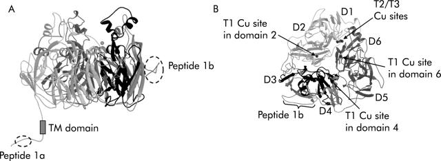
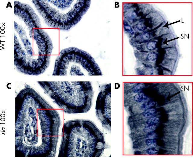
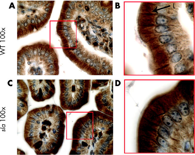
Background: Hephaestin is a multicopper ferroxidase required for basolateral transport of iron from enterocytes. Sex linked anaemia (sla) mice have a defect in the release of iron from intestinal enterocytes into the circulation due to an interstitial deletion in the hephaestin gene (heph).

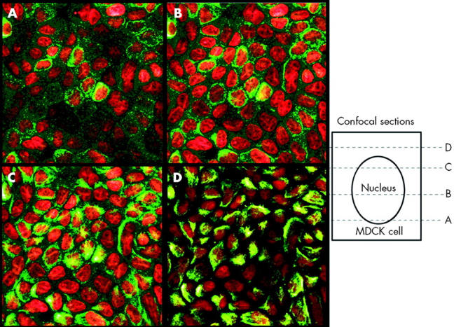
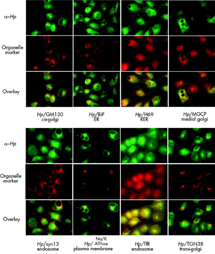
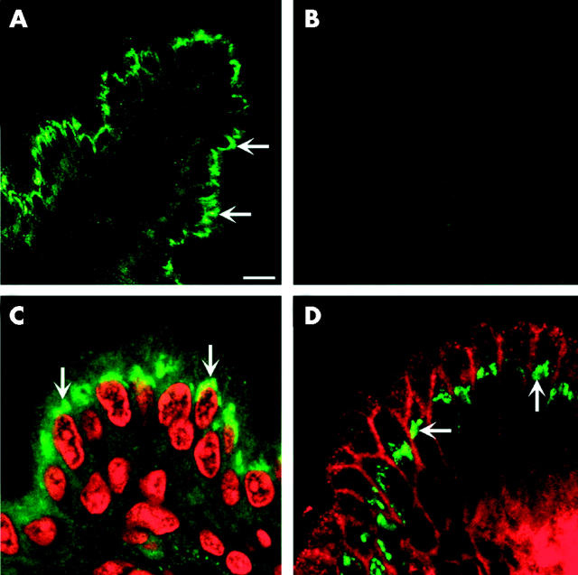
Results: We have demonstrated that hephaestin is primarily localised to a supranuclear compartment in both intestinal enterocytes and in cultured cells. In normal intestinal enterocytes, hephaestin was also present on the basolateral surface. In sla mice, hephaestin was present only in the supranuclear compartment. In contrast, the iron permease Ireg1 localised to the basolateral membrane in both control and sla mice.

Conclusion: We suggest that mislocalisation of hephaestin likely contributes to the functional defect in sla intestinal epithelium.


Figures

Figure 1
Figure 1
Location of peptide epitopes on hephaestin (Hp) and deleted region in sex linked anaemia (sla) mice. A ribbon diagram of a molecular model of mouse Hp is presented. (A) Side view of the mouse Hp molecule almost perpendicular to the pseudo-3-fold axis. Location of the two peptide epitopes (Hp peptide 1a and Hp peptide 1b) are indicated. The region deleted in sla is shown in black. (B) Top view along the pseudo-3-fold axis showing the predicted domain structure of Hp with copper binding sites indicated, the Hp1b peptide epitope, and the region deleted in sla in dark black. The figures were generated using a modified version of Molscript and subsequently rendered in Raster3D version 2.0.
Figure 2
Figure 2
Apical supranuclear location of hephaestin (Hp) in MDCK cells. Confocal immunofluorescence microscopy was carried out using an affinity purified antiserum to the C terminus of Hp in cultured MDCK cells. Hp staining is represented in green and nuclei are in red. Sequential confocal reconstructions from the bottom (A) to the top (D) of the cell showed predominantly apical perinuclear staining of Hp.
Figure 3
Figure 3
Apical supranuclear location of hephaestin (Hp) in HT29 cells. (A) Immunofluorescence microscopy using an affinity purified antiserum to the C terminus of Hp (Hp1A) in cultured HT29 cells. (B) Immunofluorescence microscopy using an affinity purified antiserum to the more N terminal part of Hp (Hp2a) in cultured HT29 cells. A single cell is shown in panels to the left and several cells are shown in the right hand panels. Hp staining is shown in green, actin in red, and nuclei in blue.
Figure 4
Figure 4
Colocalisation of hephaestin (Hp) with subcellular markers. Colocalisation of affinity purified antibodies to the C terminus of Hp (Hp1A) and antibodies to various subcellular markers in cultured CoS7 cells. Hp staining is represented in green and the markers (TfR, TGN38, GM130, BiP, H69, Na,K-ATPase, MGCP, and syntaxin 13) are shown in red. Yellow in the overlays indicates colocalisation. ER, endoplasmic reticulum; RER, rough endoplasmic reticulum; Tfr, transferrin receptor.
Figure 5
Figure 5
Predominant supranuclear location of hephaestin (Hp) in duodenal enterocytes. Immunofluorescent staining of duodenal sections from C57BL/6J mice on control diets using an antiserum to the C terminus of Hp demonstrated predominantly supranuclear staining. (A) Hp alone (green). (B) Preincubation with Hp peptide. (C) Propidium iodide staining of nucleus (in red) and Hp staining (in green). (D) Immunolocalisation of Hp (green) and Na+,K+-ATPase (red).
Figure 6
Figure 6
Absence of basolateral hephaestin (Hp) on duodenal enterocytes from sex linked anaemia (sla) mice. 3,3′-Diaminobenzidine immunohistochemistry of duodenal sections using an antiserum to the C terminus of Hp. (A) 100× of C57BL/6J (WT) duodenal sections; (B) 3× magnification of boxed area in (A). Arrows indicate lateral (L) and supranuclear (SN) staining. (C) 100× of sla duodenal sections; (D) 3× magnification of boxed area in (C). Arrows indicate supranuclear (SN) staining. There was no appreciable lateral staining.
Figure 7
Figure 7
Basolateral Ireg1 in duodenal enterocytes from sla mice. 3,3′-Diaminobenzidine immunohistochemistry of duodenal sections using an antiserum to Ireg1. (A) 100× of C57BL/6J (WT) duodenal sections; (B) 3× magnification of boxed area in (A). Arrows indicate lateral (L) staining. (C) 100× of sla duodenal sections; (D) 3× magnification of boxed area in (C).

Similar articles

Cited by

References

    1. Bothwell TH, Charlton RW, Cook JD, et al. Iron metabolism in man. Oxford: Blackwell Scientific Publications, 1979:256–83.
    1. Harris WR. Iron chemistry. In: Templeton DM, ed. Molecular and cellular iron transport. New York: Marcel Dekker Inc, 2002:1–40.
    1. Andrews NC. Animal models of iron transport and storage disorders. In: Templeton DM, ed. Molecular and cellular iron transport. New York: Marcel Dekker Inc, 2002:679–97.
    1. Johansson HE, Theil EC. Iron-response element (IRE) structure and combinatorial RNA regulation. In: Templeton DM, ed. Molecular and cellular iron transport. New York: Marcel Dekker Inc, 2002:237–53.
    1. Hanson EH, Leibold EA. Regulation of iron homeostasis by iron regulatory proteins 1 and 2. In: Templeton DM, ed. Molecular and cellular iron transport. New York: Marcel Dekker Inc, 2002:207–35.

Publication types