. 2004 Mar 16;555(Pt 3):713-25.
doi: 10.1113/jphysiol.2003.052225. Epub 2004 Jan 14.

Intrinsic cellular currents and the temporal precision of EPSP-action potential coupling in CA1 pyramidal cells

Affiliations

Intrinsic cellular currents and the temporal precision of EPSP-action potential coupling in CA1 pyramidal cells

Nikolai Axmacher et al. J Physiol. .

Abstract

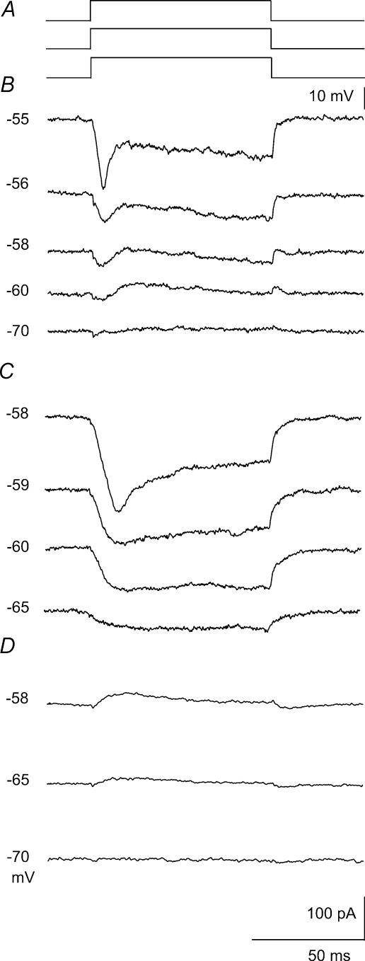
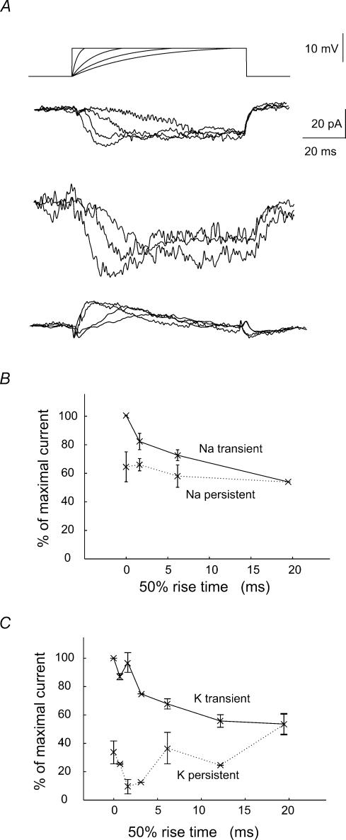
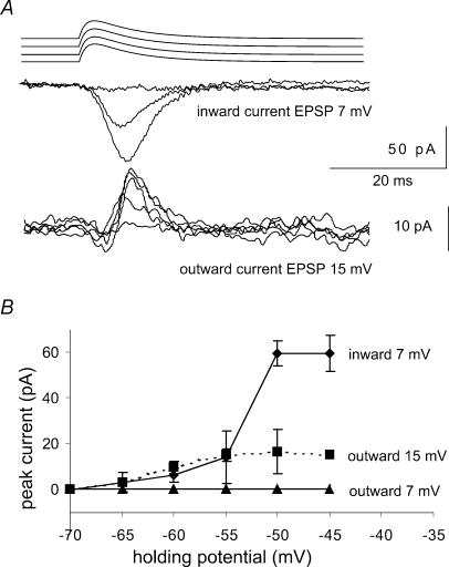
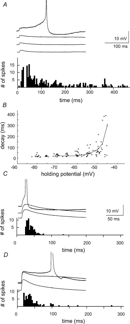
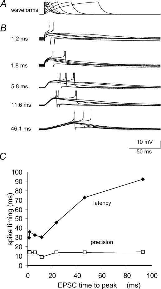
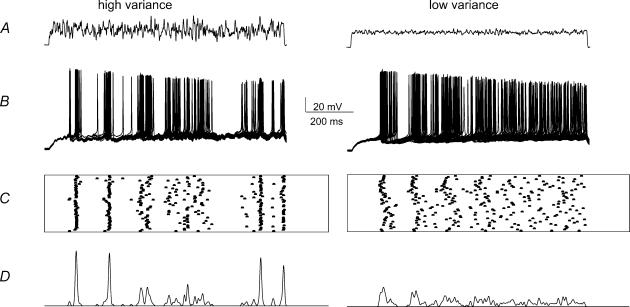
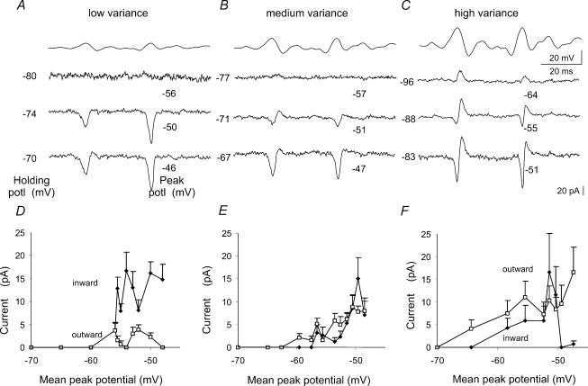
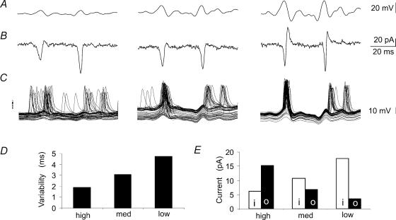
We examined relations between cellular currents activated near firing threshold and the initiation of action potentials by excitatory postsynaptic potentials (EPSPs) in CA1 pyramidal cells in vitro. Small voltage steps elicited sequences of inward-outward currents at hyperpolarized potentials, but evoked largely inward currents at near threshold potentials. Similarly small EPSP-like waveforms initiated largely inward currents while larger stimuli evoked sequences of inward followed by outward currents. Shorter rise times of EPSP-like waveforms accentuated a transient component of inward currents. Voltage clamp data were consistent with the voltage dependence of current clamp responses to injection of EPSP shaped waveforms. Small events were prolonged at subthreshold potentials and could elicit action potentials at long latencies while responses to larger EPSP waveforms showed less voltage dependence and tended to induce spikes at shorter, less variable latencies. The precision of action potentials initiated by white noise depended also on stimulus amplitude. High variance stimuli induced firing with high precision, while the timing of spikes induced by lower variance signals was more variable between trials. In voltage clamp records, high variance noise commands induced sequences of inward followed by outward currents, while lower variance versions of the same commands elicited purely inward currents. These data suggest that larger synaptic stimuli recruit outward as well as inward currents. The resulting inward-outward current sequences enhance the temporal precision of EPSP-spike coupling. Thus, CA1 pyramidal cells initiate action potentials with different temporal precision, depending on stimulus properties.


Figures

Figure 1
Figure 1. Voltage dependence of Na+ and K+ currents elicited by small current steps
A, pulse protocol: holding potential varied over the range −70 to −55mV, just below action current threshold with a standard test pulse of +10mV and duration 100ms. B, net currents elicited at different holding potentials. Note the voltage dependent switch between largely outward currents at −70 and −60mV and predominantly inward currents in a narrow range of more depolarized subthreshold potentials. C, inward currents isolated in an external solution containing 4-AP (500μm), TEA (5mm), Cs+ (2mm) and Ni2+ (100μm). D, outward currents recorded in the presence of 1μm TTX and 100μm Ni2+ (different cell). The number of cells tested was 5 for net currents, 4 for inward currents and 4 for outward currents.
Figure 2
Figure 2. Na+ and K+ currents evoked by waveforms with rising phase of differing kinetics
Pulse protocol: test waveforms consisted of an exponentially rising component of time constant 2–20ms which was then maintained at a potential depolarized by 10mV. The holding potential was −55mV and total duration of the waveform was 100ms. A shows from above the test waveforms and the net current, the pharmacologically isolated inward and the outward current. The summed inward and outward current was inward and was largely maintained during the pulse. Waveforms with the most rapid rising phase elicited a transient initial inward component. Inward current responses were recorded in the presence of 500μm 4-AP, 5mm TEA, 2mm Cs+ and 100μm Ni2+. Outward currents were recorded in the presence of TTX (1μm) and Ni2+ (100μm). B, increasing the kinetics of the rising phase of the pulse enhanced a transient component of inward currents (n= 4 cells). C, similarly, increasing the speed of the rising phase of the pulse enhanced the transient component of outward currents (n= 4 cells).
Figure 3
Figure 3. Na+ and K+ currents elicited by EPSP waveforms
Pulse protocol: holding potential varied over the range −70 to −45mV, just below action current threshold. The test pulse was an EPSP waveform of the form (1 − et/ton)et/toff with ton= 0.2ms and toff= 0.8ms and of variable peak amplitude. Inward currents were isolated in the presence of 500μm 4-AP and outward currents were examined in the presence of 1μm TTX and 100μm Ni2+. EPSP waveforms of amplitude 7mV elicited purely inward currents while larger amplitude stimuli, here 15mV, elicited both inward and outward currents. B, plot of the mean and standard deviation of inward and outward currents for EPSP waveforms of amplitudes 7 and 15mV (inward currents, n= 3 cells; outward currents n= 7 cells).
Figure 4
Figure 4. Voltage dependent amplification and firing induced by the injection of EPSP waveforms
A, the time course of potentials induced by the injection of small EPSP waveforms (amplitude 3–5mV, form (1 – et/ton)et/toff with ton= 0.2ms and toff= 0.8ms) was prolonged on membrane depolarization. Action potential initiation was examined at a holding potential where approximately 50% of trials elicited a spike. Action potentials were initiated with a latency of 125 ± 124ms (mean ±s.d.). B, the voltage dependence of the decay time constant of potentials initiated by injection of EPSP waveforms. C, the time course of potentials induced by larger EPSP waveforms (amplitude 12–16mV) changed little with holding potential. Action potentials were initiated, at a holding potential where about 50% of trials elicited a spike, with latencies in the range 11–48ms (90% of spikes). D, in the same recording, EPSPs were prolonged in the presence of 500μm 4-AP. Ninety per cent of spikes were initiated with latencies in the range of 7–96ms.
Figure 5
Figure 5. Dependence of the temporal precision of spike generation on the rise time of EPSP-like waveforms
A, current clamp stimuli followed an equation of the form (1 – et/ton)et/toff with ton= 12–90ms and toff= 0.8ms. In records from a given cell, the stimulus amplitude was adjusted to result in depolarizations of fixed amplitude close to 10mV. B, action potentials initiated by waveforms with different rise times. The holding potential was maintained at a level near −60mV, where about 50% of the stimuli initiated firing. C plots the mean latency and the standard deviation (‘precision’) of action potential latency against the time to peak of the waveforms that initiated firing (n= 6 cells).
Figure 6
Figure 6. Spike timing in response to noise stimuli of different variance
A, the noise stimuli of high and low variance. B, action potentials initiated by 50 trials using these waveforms. C, spike timing from successive trials is indicated in raster plots. D, plots of firing probability obtained by convolving the times of spike generation with a gaussian function with standard deviation equal to the duration of the trace (1 s) divided by the mean number of spikes. This procedure normalized for variations in the number of spikes.
Figure 7
Figure 7. Voltage dependence of currents elicited by noise of different variances
Pulse protocol: noise command pulses of low, medium and high variance (standard deviations of 1.8, 3.7 and 6.9mV). Holding potentials were varied between −95 and −65mV and steps of 17 and 9mV were used for the low and medium variance waveform, respectively. Extracts of responses at a range of holding potentials, indicated to the left of the trace, are shown with the peak voltage reached during the trace indicated at the right. A, the low variance noise waveform elicited purely inward currents at all holding potentials. B, noise of medium variance induced inward currents which were sometimes succeeded by small outward currents. C, as the holding potential was depolarized, high variance noise commands induced first outward currents and then sequences of inward followed by outward currents. D to F, the mean amplitude of inward and outward currents measured from 15 points corresponding to the largest depolarizing transients during each noise trace plotted against the mean peak potential for noise of low (D), medium (E) and high (F) variance. Inward currents dominate in responses to the low variance waveform, while outward currents become larger than inward currents in responses to the high variance waveform.
Figure 8
Figure 8. Membrane currents and action potentials initiated by noise stimuli
A, portions of noise waveforms with small, medium and high variance. B, resulting cellular currents. C, the timing of action potentials induced by injecting the same waveforms in current clamp experiments. The apparent action potential ‘doublets’ result from superpositions of differently timed single action potentials in distinct sweeps. D, the variability in action potential timing calculated as the mean of the standard deviation in action potential timing near points at which currents were observed in voltage clamp. Data obtained at 9–14 time points for 50 stimulus trials in records from 9 cells. E, changes in the balance of inward and outward currents, measured from voltage clamp experiments as in B, corresponding to noise stimuli of low, medium and high variance. Inward, i, and outward, o, currents were measured as shown in Fig. 7 from the most depolarized holding potential. Peak inward and outward currents were measured from the same time windows (n= 14) used to calculate variability in spike timing (n= 9 cells). Outward currents increased with the variance of the noise waveform, while the apparent amplitude of inward currents was reduced as the temporally overlapping outward currents increased in amplitude.

References

    1. Abbott LF, Nelson SB. Synaptic plasticity: taming the beast. Nat Neurosci. 2000;3(Suppl.):1178–1183. - PubMed
    1. Aksay E, Gamkrelidze G, Seung HS, Baker R, Tank DW. In vivo intracellular recording and perturbation of persistent activity in a neural integrator. Nat Neurosci. 2001;4:184–193. - PubMed
    1. Azouz R, Gray CM. Dynamic spike threshold reveals a mechanism for synaptic coincidence detection in cortical neurons in vivo. Proc Natl Acad Sci U S A. 2000;97:8110–8115. - PMC - PubMed
    1. Bolshakov VY, Siegelbaum SA. Regulation of hippocampal transmitter release during development and long-term potentiation. Science. 1995;269:1730–1734. - PubMed
    1. Brew HM, Forsythe ID. Two voltage-dependent K+ conductances with complementary functions in postsynaptic integration at a central auditory synapse. J Neurosci. 1995;15:8011–8022. - PMC - PubMed

Publication types

LinkOut - more resources