. 2004 Jan 14;24(2):514-21.
doi: 10.1523/JNEUROSCI.3408-03.2004.

The central fragment of Reelin, generated by proteolytic processing in vivo, is critical to its function during cortical plate development

Affiliations

The central fragment of Reelin, generated by proteolytic processing in vivo, is critical to its function during cortical plate development

Yves Jossin et al. J Neurosci. .

Abstract

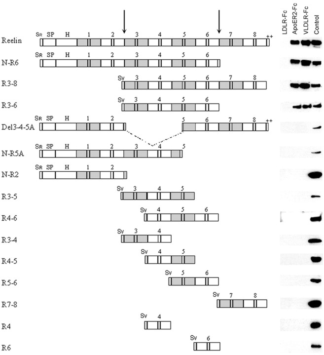
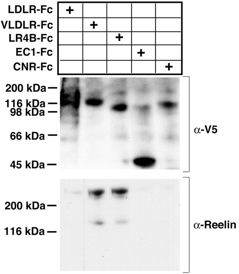
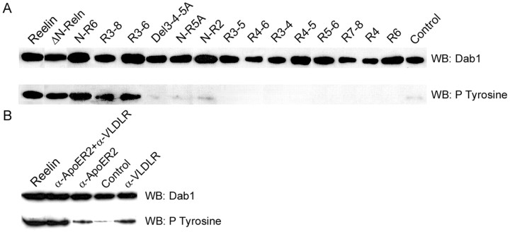
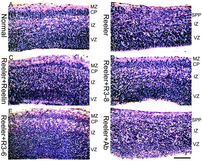
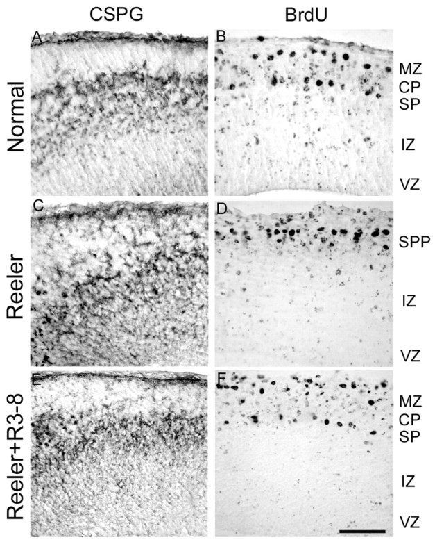
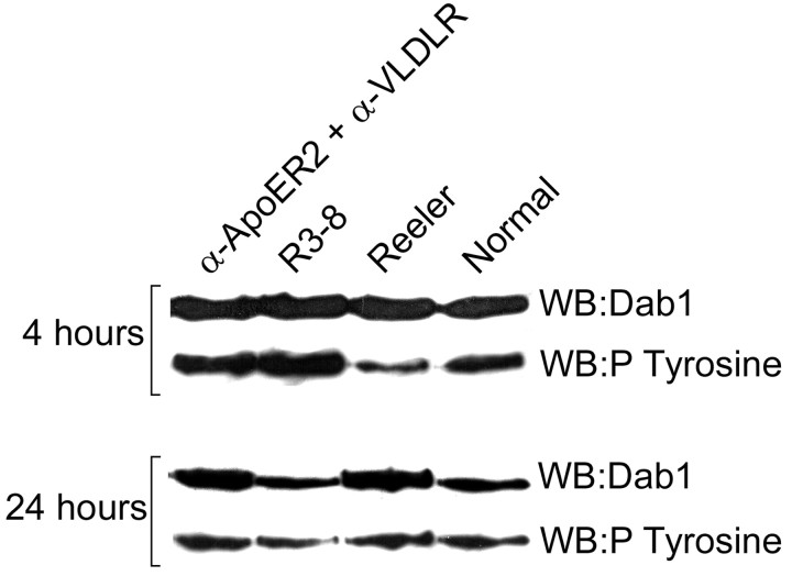
Reelin is a large extracellular protein that controls cortical development. It binds to lipoprotein receptors very-low-density lipoprotein receptor and apolipoprotein-E receptor type 2, thereby inducing phosphorylation of the adapter Dab1. In vivo, Reelin is cleaved into three fragments, but their respective function is unknown. Here we show the following: (1) the central fragment is necessary and sufficient for receptor binding in vitro and for Dab1 phosphorylation in neuronal cultures; (2) Reelin does not bind the protocadherin cadherin-related neuronal receptor (CNR1) as reported previously; (3) Reelin and its central fragment are equally able to rescue the reeler phenotype in a slice culture assay; and (4) anti-receptor antibodies can induce Dab1 phosphorylation but do not correct the reeler phenotype in slices. These observations show that the function of Reelin is critically dependent on the central fragment generated by processing but primarily independent of interactions with CNR1 and on the N-terminal region. They also indicate that events acting in parallel to Dab1 phosphorylation might be required for full activity.


Figures

Figure 1.
Figure 1.
Receptor binding. The different Reelin constructs are shown, together with their ability to coprecipitate with LDLR-Fc, ApoER2-Fc, and VLDLR-Fc. No construct binds to LDLR; only full-length Reelin and proteins N-R6, R3-6, and R3-8 are able to bind to VLDLR and ApoER2, and they bind similarly to both. The lane labeled Control is a Western blot of supernatant from cells transfected with the different plasmids. Blots were revealed with anti-Reelin G10 for the constructs that contain region H, with anti-Reelin 12 and 14 for the constructs that contain the C-terminal repeat (de Bergeyck et al., 1998), and with anti-Myc for the others. Sr, Sv, Signal peptides of Reelin and encoded by the pSecTag vector; SP; region of Reelin with similarity with F-spondin; H, unique region. Arrows indicate the two Reelin cleavage sites.
Figure 2.
Figure 2.
CNR1 does not bind Reelin. Supernatant of transfected HEK293 cells containing full-length, secreted and biologically active Reelin was incubated with the indicated Fc-fusion proteins immobilized on 30 μl of protein A-Sepharose as described in Materials and Methods. Precipitated proteins were separated by SDS gel electrophoresis and visualized by immunoblotting with the indicated antibodies. Anti-V5 monoclonal antibody equally detects all V5-tagged fusion proteins, irrespective of size. Only ApoER2 and VLDLR, and to a very small degree LDLR, bind Reelin. Neither the full ectodomain (CNR1-Fc) nor the EC1 domain fused to Fc (EC1-Fc) show any detectable Reelin binding under these conditions.
Figure 3.
Figure 3.
Dab1 phosphorylation. A, Stimulation with Reelin proteins. Primary neuronal cultures were treated for 20 min with different full-length and partial Reelin proteins (as supernatant from transfected cells) or with a control supernatant, and Dab1 was immunoprecipitated from the lysate with a rabbit polyclonal antibody against the C-terminal peptide. In the top panel, blots were revealed with a monoclonal anti-Dab1 antibody. In the bottom panel, similar blots were revealed using monoclonal anti-phosphotyrosine antibody 4G10. Only full-length ΔN-Reelin and partial N-R6, R3-6, and R3-8 Reelin proteins are able to trigger Dab1 tyrosine phosphorylation. B, Stimulation by anti-receptor antibodies. Primary neuronal cultures were treated with monoclonal antibodies, and the levels of Dab1 protein and phosphorylation were assessed as described above. Two antibodies against VLDLR and ApoER2 were found to stimulate Dab1 phosphorylation modestly when added separately but are able to induce phosphorylation to the same extent as full-length Reelin when added together. WB, Western blot.
Figure 4.
Figure 4.
Partial normalization of the reeler phenotype by Reelin, R3-8, and R3-6. Embryonic brain slices were prepared at E13.5, when no CP is present, and cultivated in vitro for 2 d. The CP that develops in vitro from a normal embryo (A) has features of the normal CP, including definition of the marginal zone (MZ) and superplate (SPP) and radial neuronal orientation. The CP that develops in a slice from a reeler embryo (B) is disorganized, flanked on its outer aspect by an SPP. A partial normalization of the reeler CP is obtained by addition of full-length (C), R3-8 (D), and R3-6 (E) recombinant Reelin but not of the anti-receptor antibody combination that is able to trigger Dab1 phosphorylation (F). Scale bar, 90 μm. IZ, Intermediate zone; Ab, antibody.
Figure 5.
Figure 5.
Partial rescue of preplate splitting in reeler slices. Preplate elements were labeled with the anti-chondroitin sulfate antibody CS-56 (A, C, E) or by BrdU injection of the pregnant mother on E10.5 (B, D, F). In normal embryos (A, B), the appearance of the CP results in preplate splitting, with preplate cells located in the marginal zone (MZ) and superplate (SP). In reeler embryos (C, D), no such splitting occurs, and all preplate cells gather in the superplate (SPP). When reeler slices are cultured in the presence of R3-8 (E, F), R3-6, or native Reelin (data not shown), a partial splitting of the preplate population is observed. Scale bar, 90 μm. IZ, Intermediate zone; CSPG, chondroitin sulfate proteoglycan.
Figure 6.
Figure 6.
Dab1 phosphorylation and protein levels in slices. reeler slices were cultured in control conditions, in the presence of active R3-8 or stimulating antibodies and compared with normal slices. Dab1 phosphorylation and protein levels were estimated after 4 and 24 hr in vitro. After 4 hr, both the antibodies and R3-8 induced a phosphorylation of Dab1 that was comparable with that in normal slices. After 24 hr, R3-8 resulted in a downregulation of Dab1 protein levels that, however, failed to occur in slices treated with antibodies.

References

    1. Andersen OM, Benhayon D, Curran T, Willnow TE (2003) Differential binding of ligands to the apolipoprotein e receptor 2. Biochemistry 42: 9355-9364. - PubMed
    1. Arnaud L, Ballif BA, Forster E, Cooper JA (2003) Fyn tyrosine kinase is a critical regulator of disabled-1 during brain development. Curr Biol 13: 9-17. - PubMed
    1. Bar I, Tissir F, Lambert De Rouvroit C, De Backer O, Goffinet AM (2003) The gene encoding disabled-1 (DAB1), the intracellular adaptor of the Reelin pathway, reveals unusual complexity in human and mouse. J Biol Chem 278: 5802-5812. - PubMed
    1. Bock HH, Herz J (2003) Reelin activates SRC family tyrosine kinases in neurons. Curr Biol 13: 18-26. - PubMed
    1. Boucher P, Liu P, Gotthardt M, Hiesberger T, Anderson RG, Herz J (2002) Platelet-derived growth factor mediates tyrosine phosphorylation of the cytoplasmic domain of the low density lipoprotein receptor-related protein in caveolae. J Biol Chem 277: 15507-15513. - PubMed

Publication types

MeSH terms

Substances

Associated data

LinkOut - more resources