. 2004 Apr 2;279(14):13478-87.
doi: 10.1074/jbc.M308523200. Epub 2004 Jan 14.

Pharmacological enhancement of beta-hexosaminidase activity in fibroblasts from adult Tay-Sachs and Sandhoff Patients

Affiliations

Pharmacological enhancement of beta-hexosaminidase activity in fibroblasts from adult Tay-Sachs and Sandhoff Patients

Michael B Tropak et al. J Biol Chem. .

Abstract

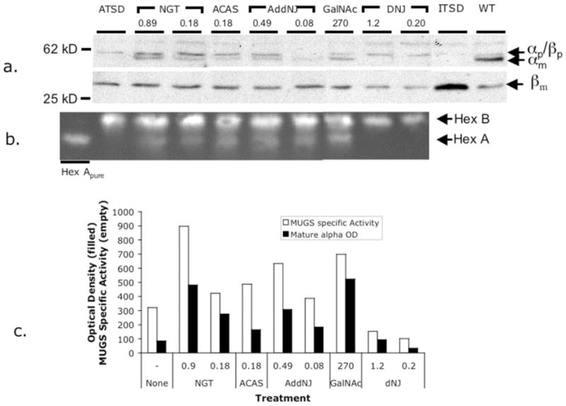
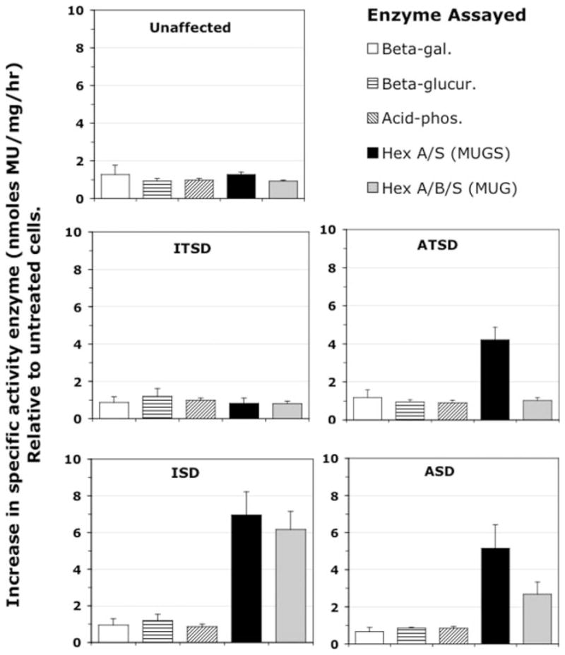
Tay-Sachs and Sandhoff diseases are lysosomal storage disorders that result from an inherited deficiency of beta-hexosaminidase A (alphabeta). Whereas the acute forms are associated with a total absence of hexosaminidase A and early death, the chronic adult forms exist with activity and protein levels of approximately 5%, and unaffected individuals have been found with only 10% of normal levels. Surprisingly, almost all disease-associated missense mutations do not affect the active site of the enzyme but, rather, inhibit its ability to obtain and/or retain its native fold in the endoplasmic reticulum, resulting in its retention and accelerated degradation. By growing adult Tay-Sachs fibroblasts in culture medium containing known inhibitors of hexosaminidase we have raised the residual protein and activity levels of intralysosomal hexosaminidase A well above the critical 10% of normal levels. A similar effect was observed in fibroblasts from an adult Sandhoff patient. We propose that these hexosaminidase inhibitors function as pharmacological chaperones, enhancing the stability of the native conformation of the enzyme, increasing the amount of hexosaminidase A capable of exiting the endoplasmic reticulum for transport to the lysosome. Therefore, pharmacological chaperones could provide a novel approach to the treatment of adult Tay-Sachs and possibly Sandhoff diseases.


Figures

Fig. 1
Fig. 1. Heat inactivation of ATSD Hex A, αGly-269 → Ser mutant, at 42 °C is attenuated in the presence of Hex inhibitors
Partially purified wild type or mutant Hex A were heated at 42 °C in the absence of inhibitors (panel a, open symbols), or in the presence of different inhibitor concentrations (filled symbols), GalNAc (panel b), ACAS (panel c), and NGT (panel d) for 5, 10, 20, 30, or 45 min. Following each incubation MUGS activity was determined. The untreated Hex A curve (panel a) is reproduced in the other panels for ease of comparison. “Fraction remaining MUGS activity” (all y-axes) was calculated using the formula (MU fluorescence of inhibitor-treated sample heated for a given time)/(MU fluorescence of inhibitor-treated sample left at 4 °C (time = 0 min), n = 3), i.e. 1 = no change.
Fig. 2
Fig. 2. Dose-dependent increase in MUGS activity following growth of ATSD fibroblasts in media containing different Hex inhibitors
ATSD fibroblasts were grown in media containing different concentrations of compounds described in Table I (identified to the right of the graph), for 5 days, lysed and MUGS activity determined. Standard deviations are shown above each data point (n = 3). The relative increases in MUGS hydrolysis were determined as (MU fluorescence of the inhibitor-treated cells)/(MU fluorescence of an untreated cells). The last data point for ACAS reflects a loss of cell viability.
Fig. 3
Fig. 3. Increased MUGS activity in treated ATSD fibroblasts is associated with an increase in the mature αsubunit protein and levels of the active Hex A isozyme
a, Western blot using a rabbit polyclonal anti-human Hex A antibody, showing increased amounts of lysosomally processed, mature β-subunits (βm) in inhibitor-treated fibroblasts. Bands corresponding to the mature β-subunits (βm) are shown in the lower strip. Position of two relevant Mr markers are shown to the left of the blot. b, resolution of the Hex isozymes from treated and untreated cell lysates by cellulose acetate electrophoresis shows increased amounts of Hex A activity (using MUG) in inhibitor treated fibroblasts (MU visualized under UV light). Bands corresponding to Hex A and Hex B are denoted by arrows to the right of the zymogram. For both a and b, the concentrations (mM) of inhibitor used are shown below the inhibitor name. c, the increase in MUGS activity parallels the increase in amount of mature α-subunit from lysates of treated ATSD fibroblasts. The specific MUGS activity (nanomoles/mg total cell protein/h) from lysates used to produce the Western blot and zymogram in a and b is shown plotted (open bars) adjacent to the optical density of the αm band (filled bars) from the Western blot in panel c. The optical density was standardized relative to the density of the bands corresponding to βm.
Fig. 4
Fig. 4. Increased MUGS (Hex A) activity is found in the lysosomal fraction from NGT treated ATSD fibroblasts
a, comparison of MUGS activity (nanomoles/mg of total cell protein/h) in the postnuclear supernatant (PNS) and lysosome-enriched (Lyso.) fraction from untreated (open bar) and NGT (0.9 mM) treated (filled bar) ATSD fibroblasts. b, Western blots comparing the levels of α-subunits of Hex (α Hex), mature β-subunits of Hex (β Hex), glucocerebrosidase (Gcase), and calnexin in the PNS and lysosomal fractions (Lyso.) from untreated and NGT-treated ATSD cells. Position of relevant Mr markers (in kDa) are shown to the left of the blots. One microgram of total protein from each of the PNS and lysosomal fractions were analyzed.
Fig. 5
Fig. 5. Kinetics of Hex A activity enhancement
a, MUGS activity in ATSD fibroblasts grown in the presence of three different concentrations of NGT over a 9-day period. Shown is the relative increase in MU fluorescence from MUGS hydrolysis (MU from treated/MU from untreated cells grown for the same length of time) in lysates from ATSD fibroblasts grown in different concentrations of NGT (corresponding symbols shown in the left-hand corner of the graph) for increasing periods of time (days). Each MUGS assay was further normalized using the acid phosphatase activity (MUP) of the same lysate. b, demonstrates that MUP hydrolysis is a valid means of normalization, because there were no changes in relative acid phosphatase activity (MUP) in another set of paired treated/untreated cell lysates over the same 9-day period. For each of the graphs, standard deviations are shown above the data points (average MU fluorescence from three wells of treated cells/three wells of untreated cells grown for the same period of time). Dashed lines denote the position at which there is no change in MU fluorescence, i.e. relative increase = 1.
Fig. 6
Fig. 6. Hex A activity persists at elevated levels for several days following removal of the inhibitors from the growth media
All panels show the relative increase in MUGS hydrolysis (MU fluorescence, see legend to Fig. 5) by Hex in lysates from ATSD fibroblasts grown in different concentrations of NGT (corresponding symbols shown in the upper left corners) for increasing periods of time (days). Initially all fibroblasts were grown in the presence of NGT (a) or ACAS (b) for 6 days. Afterward fibroblasts continued to be grown in the presence of the drug (filled symbols) or in the absence of the drugs (open symbols) following removal of the original drug containing media on the sixth day. For each of the graphs, standard deviations are shown above the data points (average time-paired values from three wells each of treated/untreated cells). Dashed lines denote the position at which there is no change in MU fluorescence, i.e. relative increase = 1. The titles to the left and bottom of all panels describe the y- and x-axis of all graphs in the figure.
Fig. 7
Fig. 7. Effect of NGT treatment on Hex activity in fibroblasts from an unaffected individual and from patients with different forms of TSD and SD
Fibroblasts from an unaffected individual (a), and patients with; ITSD (b), ISD (c), ATSD (d), or ASD (e) were grown in the presence of 4 (a–d) or 3 (e) different concentrations (mM) of NGT for 5 (a–d) or 6 days (e). The relative changes in MU fluorescence due to MUG (filled diamonds) or MUGS (filled squares) hydrolysis are shown (left-most y-axis). Each datum point (average time-paired values from four wells each of treated/untreated cells, i.e. 1 = no change) is shown along with its corresponding standard deviation. Also shown on the graphs is the MUG/MUGS ratio in treated cells (open triangles) and in untreated cells as a single open circle to the left of the other data points (the x-axis does not apply to these points), and the MUG/MUGS ratios for treated cell lysates are given on the right-most axis.
Fig. 8
Fig. 8. The effect of NGT treatment on Hex and three other lysosomal enzymes in fibroblasts from an unaffected individual, and patients with either the infantile or adult forms of TSD or SD
Relative to the change observed in MUG and MUGS hydrolysis (gray and black bars, respectively) by NGT-treated ATSD, ISD, and ASD fibroblasts, the activity of other lysosomal enzymes remains largely unaffected. Note that in ITSD cells their WT levels of Hex B (MUG/MUGS = ~150/1) are responsible for all the indicated MUGS activity. Hex B likely contributes similar amounts of MUGS activity in the ATSD and “unaffected” cell lysates. In ISD cells only Hex S is present. The average (n = 4) change in specific activity (nanomoles of MU released/mg of total cell protein/h) of Hex A/B/S (MUG (shaded bar) and MUGS (filled bar)), acid-phosphatase (diagonally hatched bar), β-glucuronidase (horizontally hatched bar), and β-galactosidase (open bar) activity was expressed relative to the corresponding values in the untreated cells, i.e. 1 = no change (x-axis). All cell lines were grown for 5 days in the absence or presence of NGT (0.9 mM).

References

    1. Gravel RA, Clarke JTR, Kaback MM, Mahuran D, Sandhoff K, Suzuki K. In: The Metabolic and Molecular Bases of Inherited Disease. 7. Scriver CR, Beaudet AL, Sly WS, Valle D, editors. Vol. 2. McGraw-Hill; New York: 1995. pp. 2839–2879.
    1. Mark BL, Mahuran DJ, Cherney MM, Zhao D, Knapp S, James MN. J Mol Biol. 2003;327:1093–1109. - PMC - PubMed
    1. Hou Y, Tse R, Mahuran DJ. Biochemistry. 1996;35:3963–3969. - PubMed
    1. Mahuran DJ. Biochim Biophys Acta. 1991;1096:87–94. - PubMed
    1. Dlott B, D’Azzo A, Quon DVK, Neufeld EF. J Biol Chem. 1990;265:17921–17927. - PubMed

Publication types

MeSH terms

Grants and funding