The cochlear cleft
- PMID: 14729522
- PMCID: PMC7974172
The cochlear cleft
Abstract
Background and purpose: Recent advances in the display of medical images permit the routine study of temporal bone CT images at high magnification. We noted an unfamiliar structure, which we now call the "cochlear cleft," in the otic capsule. To our knowledge, this report represents the first description of this structure in the medical imaging literature.
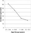
Methods: Temporal bone CT performed in 100 pediatric patients without sensorineural hearing loss were examined for the presence of cochlear clefts. Incidence of cochlear clefts as well as the relationship between age and incidence was examined.
Results: Cochlear clefts were present in 41% of the subjects. Incidence decreased with age.
Conclusion: We describe a cleft in the otic capsule that is frequently seen on magnified images of temporal bone CT studies in children. The cleft may be the fissula ante fenestram.
Figures


References
-
- Anson BJ. The labyrinths and their capsule in health and disease. Trans Am Acad Ophthalmol 1969;73:17–38 - PubMed
-
- Donaldson JA, Duckert LG, Lambert PR, Rubel EW. Anson Donaldson Surgical Anatomy of the Temporal Bone. 4th ed. New York: Raven Press;1992. :106–127
-
- Donaldson JA, Duckert LG, Lambert PR, Rubel EW. Anson Donaldson Surgical Anatomy of the Temporal Bone. 4th ed. New York: Raven Press;1992. :185
-
- Milroy CM, Michaels L. Pathology of the otic capsule. J Laryngol Otol 1990;104:83–90 - PubMed
-
- Donaldson JA, Duckert LG, Lambert PR, Rubel EW. Anson Donaldson Surgical Anatomy of the Temporal Bone. 4th ed. New York: Raven Press;1992. :430
Publication types
MeSH terms
LinkOut - more resources
Full Text Sources
Miscellaneous
