

Case Reports
Diffusion tensor MR imaging visualizes the altered hemispheric fiber connection in callosal dysgenesis
Affiliations
- PMID: 14729523
- PMCID: PMC7974170
Item in Clipboard
Case Reports
Diffusion tensor MR imaging visualizes the altered hemispheric fiber connection in callosal dysgenesis
AJNR Am J Neuroradiol.
2004 Jan.
Abstract
We report three cases of callosal dysgenesis that were evaluated by diffusion tensor imaging and fiber tractography. In partial agenesis of corpus callosum, fiber tracts from all regions of brain converged to a partially developed small genu portion and connected to the contralateral side. In complete callosal agenesis, fibers from the hemispheres failed to cross the midline and formed thick bundles running anteroposteriorly (eg, Probst bundle). The thickness of the anterior commissure was enlarged or smaller than normal brain, and other white matter tracts were not markedly different from normal brain tissue.
Figures
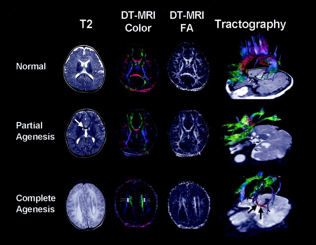
Anatomic T2-weighted images, diffusion tensor-based color maps, FA maps, and FT of callosal dysgenesis. Upper row depicts normal findings of corpus callosum; i.e., clear red fibers and high FA bundles crossing midline at diffusion tensor MR imaging. Tractography shows interhemispheric fiber connections through corpus callosum; i.e., red-colored fibers in the midline. Middle row shows partially developed corpus callosum in genu portion (white arrow). Tractography demonstrates fibers from parieto-occipital regions converging into small red-colored genu as well as fibers from frontal lobes. Fibers from the posterior part run anteriorly and form longitudinal green-colored fibers, the Probst bundle. In complete agenesis of corpus callosum, the Probst bundle is more apparent as thick, green, high-FA fibers medial to lateral ventricle (double arrows). Tractography shows thick bundles of green color consist of various fibers from ipsilateral hemisphere, Probst bundle, and thick red-colored AC (black arrows).
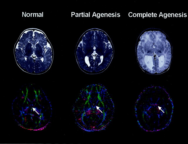
T2-weighted and diffusion tensor-based color images at the level of the AC. Right-to-left running red fibers are identified in control subject and one patient with callosal agenesis, whereas other patients did not show these transverse fascicles on diffusion tensor maps (white arrows).
Similar articles
-
Arrangement of fiber tracts forming Probst bundle in complete callosal agenesis: report of two cases with an evaluation by diffusion tensor tractography.Acta Radiol. 2006 Dec;47(10):1063-6. doi: 10.1080/02841850600930025. Acta Radiol. 2006. PMID: 17135009
-
Assessing prenatal white matter connectivity in commissural agenesis.Brain. 2013 Jan;136(Pt 1):168-79. doi: 10.1093/brain/aws332. Brain. 2013. PMID: 23365096
-
Organising white matter in a brain without corpus callosum fibres.Cortex. 2015 Feb;63:155-71. doi: 10.1016/j.cortex.2014.08.022. Epub 2014 Sep 11. Cortex. 2015. PMID: 25282054
-
[Agenesis of the corpus callosum].Radiologe. 2018 Jul;58(7):636-645. doi: 10.1007/s00117-018-0388-2. Radiologe. 2018. PMID: 29774379 Review. German.
-
[Congenital malformations of the brain. 2: Malformations of the corpus callosum and holoprocencephalies].Radiologe. 2003 Nov;43(11):925-33. doi: 10.1007/s00117-003-0975-7. Radiologe. 2003. PMID: 14628116 Review. German.
Cited by
-
The use of diffusion tractography to characterize a corpus callosum malformation in a dog.J Vet Intern Med. 2019 Mar;33(2):743-750. doi: 10.1111/jvim.15392. Epub 2018 Dec 26. J Vet Intern Med. 2019. PMID: 30588678 Free PMC article.
-
Variability of homotopic and heterotopic callosal connectivity in partial agenesis of the corpus callosum: a 3T diffusion tensor imaging and Q-ball tractography study.AJNR Am J Neuroradiol. 2009 Feb;30(2):282-9. doi: 10.3174/ajnr.A1361. Epub 2008 Nov 11. AJNR Am J Neuroradiol. 2009. PMID: 19001538 Free PMC article.
-
Shared microstructural features of behavioral and substance addictions revealed in areas of crossing fibers.Biol Psychiatry Cogn Neurosci Neuroimaging. 2017 Mar;2(2):188-195. doi: 10.1016/j.bpsc.2016.03.001. Biol Psychiatry Cogn Neurosci Neuroimaging. 2017. PMID: 28367515 Free PMC article.
-
Quantitative brain morphological analysis in CHARGE syndrome.Neuroimage Clin. 2019;23:101866. doi: 10.1016/j.nicl.2019.101866. Epub 2019 May 21. Neuroimage Clin. 2019. PMID: 31154243 Free PMC article.
-
Plasticity of Interhemispheric Temporal Lobe White Matter Pathways Due to Early Disruption of Corpus Callosum Development in Spina Bifida.Brain Connect. 2016 Apr;6(3):238-48. doi: 10.1089/brain.2015.0387. Epub 2016 Jan 22. Brain Connect. 2016. PMID: 26798959 Free PMC article.
References
-
- Probst M. Über den Bau des vollständing balkenlosen Groβhirns. Arch Psychiatr 1901;34:709–786
-
- Barkovich AJ, Norman D. Anomalies of the corpus callosum: correlation with further anomalies of the brain. AJNR Am J Neuroradiol 1988;9:493–501 - PubMed
-
- Mori S, Crain BJ, Chacko VP, van Zijl PCM. Three dimensional tracking of axonal projections in the brain by magnetic resonance imaging. Ann Neurol 1999;45:265–269 - PubMed
Publication types
MeSH terms
LinkOut - more resources
Full Text Sources