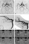
Improved image quality of intracranial aneurysms: 3.0-T versus 1.5-T time-of-flight MR angiography
- PMID: 14729534
- PMCID: PMC7974183
Improved image quality of intracranial aneurysms: 3.0-T versus 1.5-T time-of-flight MR angiography
Abstract
Background and purpose: We hypothesize that the nearly doubling of signal-to-noise ratio at 3.0 T compared with that at 1.5 T yields improved clinical MR angiograms and enables superior visualization of intracranial aneurysms. The goal of this study was to determine whether 3.0-T time-of-flight (TOF) MR angiography is superior to 1.5-T TOF MR angiography in the detection and characterization of intracranial aneurysms.
Methods: Fifty consecutive patients referred for MR angiography of a known or suspected intracranial aneurysm underwent 3-T TOF MR angiography. Seventeen of these 50 patients had also previously undergone 1.5-T TOF MR angiography and these images were used as a basis for comparison with images obtained at 3.0 T. Fourteen of 23 patients in whom aneurysms were identified also underwent prior conventional angiography, which was used as the reference standard. Readers blinded to patient history identified the presence and location of aneurysm(s) on angiograms and graded images for overall image quality by using a five-point scale.
Results: Twenty-eight aneurysms were identified in 23 of 50 patients. Seventeen aneurysms in 17 patients had been documented with 1.5-T MR angiography. The 3.0-T technique had a higher mean image quality score than that of the 1.5-T MR technique (P <.0001). Both 3.0-T and 1.5-T TOF MR angiography depicted all the aneurysms that had been documented by conventional angiography.
Conclusion: 3D TOF MR angiography at 3 T offers superior depiction of intracranial aneurysms compared with that of 1.5-T TOF MR angiography.
Figures


References
-
- Menghini VV, Brown RD Jr, Sicks JD, et al. Incidence and prevalence of intracranial aneurysms and hemorrhage in Olmsted County, Minnesota, 1965 to 1995. Neurology 1998;51:405–411 - PubMed
-
- International Study of Unruptured Intracranial Aneurysms Investigators. Unruptured intracranial aneurysms: natural history, clinical outcome, and risks of surgical and endovascular treatment. Lancet 2003;362:103–110 - PubMed
-
- Fogelholm R, Hernesniemi J, Vapalahti M. Impact of early surgery on outcome after aneurysmal subarachnoid hemorrhage: a population-based study. Stroke 1993;24:1649–1654 - PubMed
-
- Huston J III, Rufenacht DA, Ehman RL, et al. Intracranial aneurysms and vascular malformations: comparison of time-of-flight and phase-contrast MR angiography. Radiology 1991;181:721–730 - PubMed
Publication types
MeSH terms
Grants and funding
LinkOut - more resources
Full Text Sources
Medical
