

The sphenoparietal sinus of breschet: does it exist? An anatomic study
- PMID: 14729539
- PMCID: PMC7974157
The sphenoparietal sinus of breschet: does it exist? An anatomic study
Erratum in
- AJNR Am J Neuroradiol. 2004 Mar;25(3):B1
Abstract
Background and purpose: The termination of the superficial middle cerebral vein is classically assimilated to the sphenoid portion of the sphenoparietal sinus. This notion has, however, been challenged in a sometimes confusing literature. The purpose of the present study was to evaluate the actual anatomic relationship existing between the sphenoparietal sinus and the superficial middle cerebral vein.
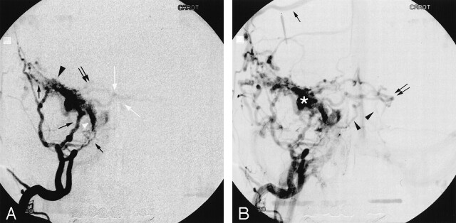
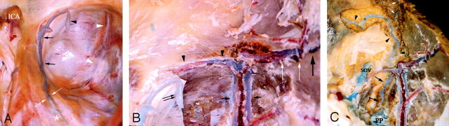
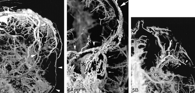
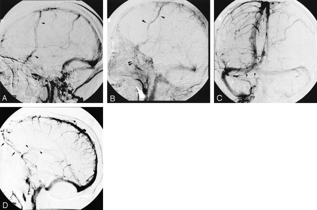
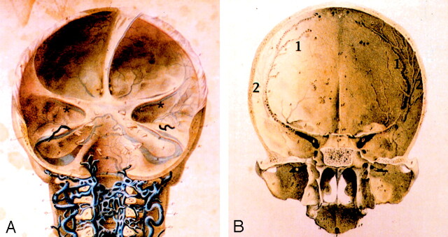
Methods: The cranial venous system of 15 nonfixed human specimens was evaluated by the corrosion cast technique (12 cases) and by classic anatomic dissection (three cases). Angiographic correlation was provided by use of the digital subtraction technique.
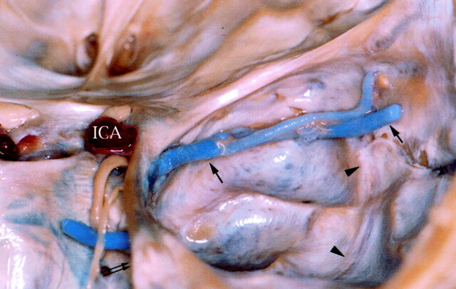
Results: The parietal portion of the sphenoparietal sinus was found to correspond to the parietal portion of the anterior branch of the middle meningeal veins. The sphenoid portion of the sphenoparietal sinus was found to be an independent venous sinus coursing under the lesser sphenoid wing, the sinus of the lesser sphenoid wing, which was connected medially to the cavernous sinus and laterally to the anterior middle meningeal veins. The superficial middle cerebral vein drained into a paracavernous sinus, a laterocavernous sinus, or a cavernous sinus but was never connected to the sphenoparietal sinus. All these venous structures were demonstrated angiographically.
Conclusion: The sphenoparietal sinus corresponds to the artificial combination of two venous structures, the parietal portion of the anterior branch of the middle meningeal veins and a dural channel located under the lesser sphenoid wing, the sinus of the lesser sphenoid wing. The classic notion that the superficial middle cerebral vein drains into or is partially equivalent to the sphenoparietal sinus is erroneous. Our study showed these structures to be independent of each other; we found no instance in which the superficial middle cerebral vein was connected to the anterior branch of the middle meningeal veins or the sinus of the lesser sphenoid wing. The clinical implications of these anatomic findings are discussed in relation to dural arteriovenous fistulas in the region of the lesser sphenoid wing.
Figures
References
-
- Breschet G. Recherches Anatomiques,Physiologiques et Pathologiques sur le Système Veineux et Spécialement sur les Canaux Veineux des Os. Paris: Villeret et Rouen;1829. :1–42
-
- Cruveilhier J. Traité d’anatomie Descriptive: Angéologie. 3rd ed. Paris: Labé;1852. :43
-
- Rouvière H, Delmas A. Anatomie humaine. Tome I: Tête et Cou. 14th ed Paris: Masson;1997;233
-
- Galligioni F, Bernardi R, Pellone M, et al. The superficial sylvian vein in normal and pathologic cerebral angiography. Am J Roentgenol Radium Ther Nucl Med 1969;107:565–578 - PubMed
-
- Hédon CE. Etude anatomique sur la circulation veineuse de l’encéphale. Thèse de la Faculté de Médecine de Bordeaux;1888. :1–96
Publication types
MeSH terms
LinkOut - more resources
Full Text Sources
Miscellaneous