. 2004 Jan 15;18(2):126-31.
doi: 10.1101/gad.1165104. Epub 2004 Jan 16.

p63 is the molecular switch for initiation of an epithelial stratification program

Affiliations

p63 is the molecular switch for initiation of an epithelial stratification program

Maranke I Koster et al. Genes Dev. .

Abstract

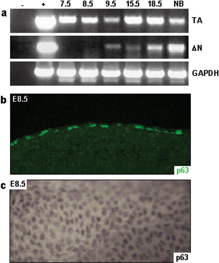
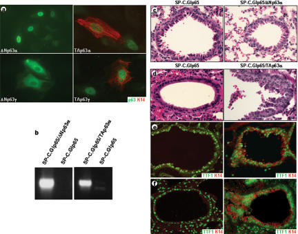
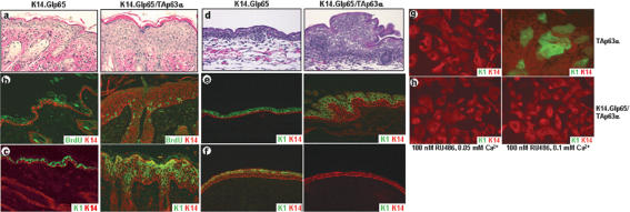
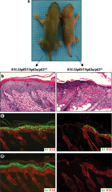
Development of stratified epithelia, such as the epidermis, requires p63 expression. The p63 gene encodes isoforms that contain (TA) or lack (DeltaN) a transactivation domain. We demonstrate that TAp63 isoforms are the first to be expressed during embryogenesis and are required for initiation of epithelial stratification. In addition, TAp63 isoforms inhibit terminal differentiation, suggesting that TAp63 isoforms must be counterbalanced by DeltaNp63 isoforms to allow cells to respond to signals required for maturation of embryonic epidermis. Our data demonstrate that p63 plays a dual role: initiating epithelial stratification during development and maintaining proliferative potential of basal keratinocytes in mature epidermis.


Figures

Figure 1.
Figure 1.
Primary p63-/- surface epithelial cells are blocked in their commitment to a stratified epithelial lineage. Differentiation markers K5 and K14, which are expressed in epithelia that have committed to a stratification program, are not expressed in primary p63-/- cells. These cells do, however, express K18, a marker for single-layered epithelia.
Figure 2.
Figure 2.
Differential expression of p63 isoforms occurs during embryonic development. RT-PCR analysis using primers specific for TA- or ΔNp63 demonstrated that TAp63 isoforms are expressed as early as E7.5, whereas ΔNp63 isoforms are not expressed until E9.5 (a). Immunofluorescence (b) and whole-mount immunohistochemistry (c) using the mAb4A4 antibody confirmed nuclear localization of TAp63 isoforms in the surface ectoderm of E8.5 embryos.
Figure 3.
Figure 3.
Ectopic expression of TAp63α initiates a stratification program in vitro and in vivo. Immunofluorescence on Ptk2 cells transfected with the indicated p63 expression constructs demonstrated that only TAp63 isoforms could induce K14 expression (∼75% of the transfected cells; a). To determine if p63 could also induce a stratification program in vivo, we induced ectopic expression of TAp63α or ΔNp63α in single-layered lung epithelia by using gene-switch mice. Induction of TAp63α or ΔNp63α transgene expression was determined by RT-PCR (b). Ectopic expression of ΔNp63α in single-layered lung epithelia did not alter lung morphology (c). Ectopic expression of TAp63α, however, resulted in squamous metaplastic lesions in the bronchioles (d) and induction of K14 expression, a marker for stratified epithelia, in adult (e) and E18.5 (f) lungs. TTF1 staining was used to identify type II alveolar cells and distal Clara cells in the bronchioles.
Figure 4.
Figure 4.
Induction of TAp63α expression results in hyperplasia, hyperproliferation, and inhibition of terminal differentiation. Induction of TAp63α expression in adult skin resulted in hyperplasia (a), hyperproliferation as demonstrated by a BrdU incorporation assay (b), and a delayed onset of terminal differentiation as demonstrated by a delayed onset of K1 expression (c). Induction of TAp63α at E8.5 resulted in hyperplasia (d) and a delayed onset of K1 expression (e) at E18.5. When TAp63α expression was induced starting at E3.5, at E13.5 the developing epidermis covering the fore-and hindlimbs demonstrated a lack of K1 expression (f). The ability of TAp63α to block terminal differentiation was further demonstrated by the failure of primary keratinocytes, in which TAp63α expression was induced, to undergo Ca2+-induced differentiation (g,h).
Figure 5.
Figure 5.
Induction of TAp63α expression in the presence of reduced levels of ΔNp63α results in epidermal fragility. TAp63α expression was induced in the epidermis of K14.Glp65/TAp63α mice on a p63+/- or p63+/+ genetic background. Although K14.Glp65/TAp63α/p63+/+ mice looked macroscopically normal after 4 d of RU486 treatment, K14.Glp65/TAp63α/p63+/- mice developed fissures in the epidermis (a). Histologically, the lesions in K14.Glp65/TAp63α/p63+/- epidermis appeared eroded (b–d). In the epidermis adjacent to these lesions, K1 expression was confined to the outermost layers of the epidermis (c), and loricrin expression was absent (d), providing an explanation for the apparent fragility of the epidermis.

Comment in

References

    1. Berton T.R., Wang, X.J., Zhou, Z., Kellendonk, C., Schutz, G., Tsai, S., and Roop, D.R. 2000. Characterization of an inducible, epidermal-specific knockout system: Differential expression of lacZ in different Cre reporter mouse strains. Genesis 26: 160-161. - PubMed
    1. Byrne C., Tainsky, M., and Fuchs, E. 1994. Programming gene expression in developing epidermis. Development 120: 2369-2383. - PubMed
    1. Cao T., He, W., Roop, D.R., and Wang, X.J. 2002. K14-GLp65 transactivator induces transgene expression in embryonic epidermis. Genesis 32: 189-190. - PubMed
    1. Dohn M., Zhang, S., and Chen, X. 2001. p63alpha and DeltaNp63alpha can induce cell cycle arrest and apoptosis and differentially regulate p53 target genes. Oncogene 20: 3193-3205. - PubMed
    1. Ghioni P., Bolognese, F., Duijf, P.H., van Bokhoven, H., Mantovani, R., and Guerrini, L. 2002. Complex transcriptional effects of p63 isoforms: Identification of novel activation and repression domains. Mol.Cell. Biol. 22: 8659-8668. - PMC - PubMed

Publication types