. 2004 Feb;186(3):638-45.
doi: 10.1128/JB.186.3.638-645.2004.

The Streptococcus gordonii platelet binding protein GspB undergoes glycosylation independently of export

Affiliations

The Streptococcus gordonii platelet binding protein GspB undergoes glycosylation independently of export

Barbara A Bensing et al. J Bacteriol. 2004 Feb.

Abstract

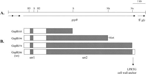
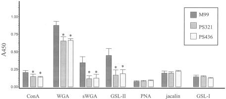
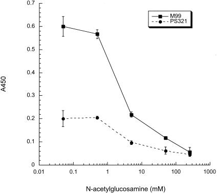
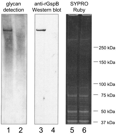
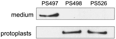
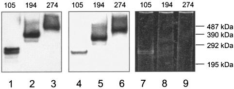
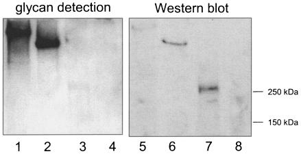
The binding of bacteria and platelets may play a central role in the pathogenesis of infective endocarditis. Platelet binding by Streptococcus gordonii strain M99 is predominantly mediated by the 286-kDa cell wall-anchored protein GspB. This unusually large protein lacks a typical amino-terminal signal peptide and is translocated from the cytoplasm via a dedicated transport system. A 14-kb segment just downstream of gspB encodes SecA2 and SecY2, two components of the GspB-specific transport system. The downstream segment also encodes several putative glycosyl transferases that may be responsible for the posttranslational modification of GspB. In this study, we compared the abilities of M99 and two GspB(-) mutant strains to bind various lectins. GspB was found to have affinity for lectins that bind N-acetylglucosamine. We also examined variant forms of GspB that lack a carboxy-terminal cell wall-anchoring domain and thus are free of covalent linkage to cell wall peptidoglycan. Like native GspB, these truncated proteins appear to be heavily glycosylated, as evidenced by migration during sodium dodecyl sulfate-polyacrylamide gel electrophoresis with an apparent molecular mass >100 kDa in excess of the predicted mass, negligible staining with conventional protein stains, and reactivity with hydrazide following periodate oxidation. Furthermore, analysis of the carbohydrate associated with the GspB variants by high-pH anion-exchange chromatography revealed the presence of approximately 70 to 100 monosaccharide residues per GspB polypeptide (primarily N-acetylglucosamine and glucose). Analysis of GspB in protoplasts of secA2 or secY2 mutant strains, which do not export GspB, indicates that GspB is glycosylated in the cytoplasm of these strains. The combined data suggest that the native GspB is a glycoprotein and that it may be glycosylated prior to export.


Figures

FIG. 1.
FIG. 1.
gspB-sec locus of M99. GspB is a cell surface-anchored platelet binding adhesin. Gly, Nss, and Gtf are likely to function in carbohydrate metabolism: Gly is predicted to be a cytoplasmic glycosyl transferase (family 8); Nss is similar to nucleotide sugar synthetases; and Gtf is 46% similar to the Bacillus subtilis polyglycerol phosphate α-glucosyl transferase (S06048). SecA2 and SecY2 are similar to the SecA ATPases and the SecY transmembrane translocases of various organisms (components of the general secretory pathway), respectively, and are required specifically for the export of GspB. The proteins encoded by orf1 to orf4 show no similarity to any proteins of known function.
FIG. 2.
FIG. 2.
Features of the serine-rich repeat protein GspB and the corresponding genetic locus. (A) Restriction map of the M99 chromosomal region spanning gspB. The locations of selected restriction sites in gspB are indicated. H3, HindIII; H2, HincII; Nh, NheI; Ns, NsiI; S, SpeI. (B) Diagram of GspB and truncated derivatives. The number associated with the GspB designation indicates the predicted molecular mass in kilodaltons. GspB194 was engineered with a C-terminal His6 tag. The hatched sections correspond to srr1 and srr2. wt, wild type.
FIG. 3.
FIG. 3.
Lectin binding to S. gordonii strain M99 and the GspB strains PS321 and PS436 (left to right). Biotinylated forms of the indicated lectins were assessed for binding to bacteria that were immobilized in 96-well plates. The asterisks indicate values that are significantly different from that of the parental strain, M99 (P < 0.05). The error bars indicate standard deviations.
FIG. 4.
FIG. 4.
Inhibition of succinylated-WGA binding to M99 by GlcNAc. The effect of GlcNAc on the binding of succinylated WGA to M99 and the GspB strain PS321 was assessed by the whole-cell lectin binding assay.
FIG. 5.
FIG. 5.
Detection of carbohydrate linked to GspB and other S. gordonii cell wall proteins. The proteins extracted from M99 (lanes 1, 3, and 5) or the gspB mutant strain PS436 (lanes 2, 4, and 6) were separated by electrophoresis through 3 to 8% polyacrylamide gradient gels. The proteins were then transferred to nitrocellulose and either examined for the presence of carbohydrate by using the DIG-glycan detection kit (lanes 1 and 2) or probed with a polyclonal anti-rGspB serum (lanes 3 and 4). To confirm that comparable amounts of total protein were loaded in each lane, gels run in parallel were stained with SYPRO Ruby (lanes 5 and 6).
FIG. 6.
FIG. 6.
Dependence of GspB194 export on components of the accessory Sec system. GspB194 was detected using an anti-His6 monoclonal antibody. The upper lanes contain proteins precipitated from 160 μl of the culture medium; the lower lanes were loaded with protoplasts of bacteria in 120 μl of culture. PS498 and PS526 are derivatives of strain PS497 that have mutations in secY2 and secA2, respectively.
FIG. 7.
FIG. 7.
SDS-PAGE analyses of truncated derivatives of GspB. The proteins were purified from S. gordonii culture supernatants. The predicted masses (in kilodaltons) of the GspB variants are indicated along the top of the gel. The proteins in lanes 1 to 3 (1 pmol of GspB per lane) underwent Western blot analysis using a polyclonal anti-rGspB serum. The proteins in lanes 4 to 6 (1 pmol per lane) were analyzed for the presence of carbohydrate, using the DIG-glycan detection kit. The proteins in lanes 7 to 9 (4 pmol per lane) were stained with SYPRO Ruby. Note that the proteins in lanes 8 and 9 are refractory to staining with SYPRO Ruby and are thus not readily apparent here. The molecular mass standards correspond to cross-linked multimers of phosphorylase b. Lanes 1, 4, and 7, GspB105; lanes 2, 5, and 8, GspB194; lanes 3, 6, and 9, GspB274.
FIG. 8.
FIG. 8.
Detection of glycosylated GspB in S. gordonii protoplasts. Each lane contained material from protoplasts of cells in a 120-μl culture volume. Lanes 1 to 4, proteins were examined for carbohydrate. Lanes 5 to 8, proteins were subjected to Western blot analysis using the anti-rGspB serum. Lanes 1 and 5, PS426 (GspB286 SecY2); lanes 2 and 6, PS498 (GspB194 SecY2); lanes 3 and 7, PS466 (GspB105 SecY2); lanes 4 and 8, PS436 (GspB).

References

    1. Ausubel, F. M., R. Brent, R. E. Kingston, D. D. Moore, J. G. Seidman, J. A. Smith, and K. Struhl. 1997. Current protocols in molecular biology. John Wiley & Sons, New York, N.Y.
    1. Bensing, B. A., and P. M. Sullam. 2002. An accessory sec locus of Streptococcus gordonii is required for export of the surface protein GspB and for normal levels of binding to human platelets. Mol. Microbiol. 44:1081-1094. - PubMed
    1. Benz, I., and M. A. Schmidt. 2002. Never say never again: protein glycosylation in pathogenic bacteria. Mol. Microbiol. 45:267-276. - PubMed
    1. Claverys, J. P., A. Dintilhac, E. V. Pestova, B. Martin, and D. A. Morrison. 1995. Construction and evaluation of new drug-resistance cassettes for gene disruption mutagenesis in Streptococcus pneumoniae, using an ami test platform. Gene 164:123-128. - PubMed
    1. Durack, D. T. 1975. Experimental bacterial endocarditis. IV. Structure and evolution of very early lesions. J. Pathol. 115:81-89. - PubMed

Publication types