. 2004 Feb;16(2):465-77.
doi: 10.1105/tpc.016907. Epub 2004 Jan 16.

The Arabidopsis thaliana dihydroxyacetone phosphate reductase gene SUPPRESSSOR OF FATTY ACID DESATURASE DEFICIENCY1 is required for glycerolipid metabolism and for the activation of systemic acquired resistance

Affiliations

The Arabidopsis thaliana dihydroxyacetone phosphate reductase gene SUPPRESSSOR OF FATTY ACID DESATURASE DEFICIENCY1 is required for glycerolipid metabolism and for the activation of systemic acquired resistance

Ashis Nandi et al. Plant Cell. 2004 Feb.

Abstract

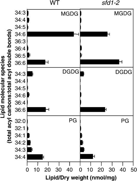
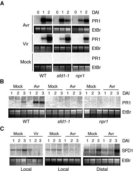
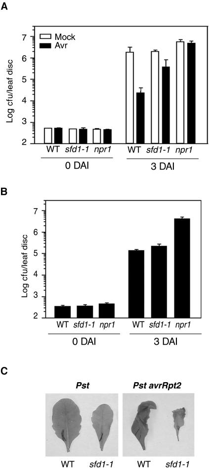
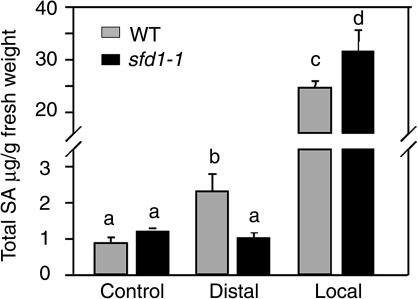
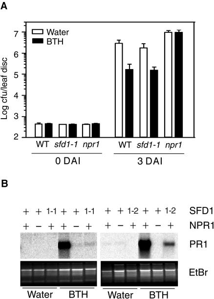
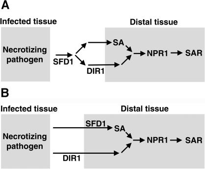
Systemic acquired resistance (SAR) is a broad-spectrum resistance mechanism in plants that is activated in naive organs after exposure of another organ to a necrotizing pathogen. The organs manifesting SAR exhibit an increase in levels of salicylic acid (SA) and expression of the PATHOGENESIS-RELATED1 (PR1) gene. SA signaling is required for the manifestation of SAR. We demonstrate here that the Arabidopsis thaliana suppressor of fatty acid desaturase deficiency1 (sfd1) mutation compromises the SAR-conferred enhanced resistance to Pseudomonas syringae pv maculicola. In addition, the sfd1 mutation diminished the SAR-associated accumulation of elevated levels of SA and PR1 gene transcript in the distal leaves of plants previously exposed to an avirulent pathogen. However, the basal resistance to virulent and avirulent strains of P. syringae and the accumulation of elevated levels of SA and PR1 gene transcript in the pathogen-inoculated leaves of sfd1 were not compromised. Furthermore, the application of the SA functional analog benzothiadiazole enhanced disease resistance in the sfd1 mutant plants. SFD1 encodes a putative dihydroxyacetone phosphate (DHAP) reductase, which complemented the glycerol-3-phosphate auxotrophy of the DHAP reductase-deficient Escherichia coli gpsA mutant. Plastid glycerolipid composition was altered in the sfd1 mutant plant, suggesting that SFD1 is involved in lipid metabolism and that an SFD1 product lipid(s) is important for the activation of SAR.


Figures

Figure 1.
Figure 1.
Major Plastidic Glycerolipids in Leaves of Wild-Type and sfd1-2 Mutant Plants. Individual glycerolipids are characterized by their head groups, MGDG, DGDG, and PG, and the total number of acyl carbons and double bonds. Lipid concentrations are expressed as nanomoles of lipids per milligram of dry weight of leaf tissue. All values are the mean of five samples ± sd. See Figure 1 in the supplemental data online for a comparison of the composition of nonplastidic glycerolipids in wild-type and sfd1-2 mutant plants.
Figure 2.
Figure 2.
PR1 and SFD1 Gene Transcript Accumulation in Pathogen-Challenged and Distal Leaves of Wild-Type and Mutant Plants. (A) PR1 gene transcript accumulation in pathogen-inoculated leaves. PR1 transcript accumulation was monitored at 0, 1, and 2 DAI in the pathogen-inoculated leaves of wild-type, sfd1-1, and npr1-5 (npr1) plants. Four fully expanded leaves of each plant were inoculated with a suspension containing 2 × 10⁵ cfu/mL of the avirulent strain P. s. tomato DC3000 carrying avrRpt2 (Avr) or the virulent strain P. s. maculicola (Vir) in 10 mM MgCl2. As a control, in parallel, RNA also was extracted from leaves treated with 10 mM MgCl2 (Mock). The blots were hybridized with a radiolabeled PR1 probe. (B) PR1 gene transcript accumulation accompanying the activation of SAR. PR1 transcript accumulation in the distal uninoculated leaves of wild-type, sfd1-1, and npr1 plants that had two other leaves previously inoculated either with a suspension containing 107 cfu/mL of P. s. tomato DC3000 carrying avrRpt2 (Avr) or 10 mM MgCl2 (Mock). RNA was extracted at 1, 2, and 3 DAI. The blot was hybridized with a radiolabeled PR1 probe. (C) SFD1 gene transcript accumulation in leaves of the wild-type plant. SFD1 transcript accumulation was monitored in leaves infiltrated with 10 mM MgCl2 (Mock; Local) and in leaves inoculated with 2 × 10⁵ cfu/mL of P. s. maculicola (Vir; Local) and P. s. tomato DC3000 carrying avrRpt2 (Avr; Local). SFD1 transcript accumulation also was monitored in the uninoculated leaves (Distal) of plants that had two other leaves infiltrated with 10 mM MgCl2 (Mock; Distal) or 2 × 10⁵ cfu/mL of P. s. tomato DC3000 carrying avrRpt2 (Avr; Distal). Blots were hybridized with a radiolabeled SFD1 probe. In (A), (B), and (C), gel loading was monitored by photographing the ethidium bromide (EtBr)–stained gel before transfer to the membrane.
Figure 3.
Figure 3.
Pathogen Growth and HR in Leaves of Wild-Type and Mutant Plants. (A) Growth of the virulent pathogen P. s. maculicola with or without the prior induction of SAR. Two fully expanded leaves of wild-type, sfd1-1, and npr1-5 (npr1) plants either were injected with 10 mM MgCl2 (Mock) (open bar) or inoculated with a suspension containing 107 cfu/mL of P. s. tomato DC3000 carrying the avirulence gene avrRpt2 (Avr) (black bar). Two days later, four other leaves of each plant were challenge-inoculated with a suspension containing 2.5 × 10⁵ cfu/mL of P. s. maculicola. P. s. maculicola numbers in leaves were monitored on the day of challenge inoculation (0 DAI) and 3 DAI. All values are the mean of five samples ±sd. (B) Growth of the avirulent pathogen P. s. tomato DC3000 containing avrRpt2. Four fully expanded leaves of wild-type, sfd1-1, and npr1 plants were injected with a suspension containing 2 × 10⁵ cfu/mL of P. s. tomato DC3000 carrying the avirulence gene avrRpt2. Bacterial growth in the pathogen-inoculated leaves was monitored at 0 and 3 DAI. All values are the mean of five samples ±sd. (C) HR in leaves of the wild-type and sfd1-1 mutant. Fully expanded leaves of the wild-type and the sfd1-1 mutant were inoculated with a suspension containing 107 cfu/mL of P. s. tomato carrying the avirulence gene avrRpt2 (Pst avrRpt2). As a control, in parallel, leaves from the wild-type and sfd1-1 plants also were inoculated with the virulent strain P. s. tomato (Pst). The HR-associated tissue collapse was monitored over a period of 24 h. The photographs shown were taken at the same magnification, 16 h after inoculation with the pathogen.
Figure 4.
Figure 4.
Total SA Content in the Leaves of Pathogen-Inoculated Wild-Type and sfd1-1 Mutant Plants. Total SA (SA + SAG) accumulation in the wild-type (gray bars) and sfd1-1 (black bars) plants. Leaf samples were taken from untreated plants (Control) and from the pathogen-inoculated (Local) and uninoculated leaves (Distal) of a plant inoculated 2 d earlier with a suspension containing 107 cfu/mL of P. s. tomato DC3000 carrying the avirulence gene avrRpt2. The total SA concentration is expressed as micrograms of total SA per gram of fresh weight of leaf tissue. All values are the mean of five samples ±sd. The difference between any two bars with different letters at the top is statistically significant (>95% confidence), whereas bars with the same letter indicates that the difference between the two values is statistically insignificant.
Figure 5.
Figure 5.
Growth of P. s. maculicola and PR1 Transcript Accumulation in BTH-Treated Leaves of Wild-Type and Mutant Plants. (A) Growth of P. s. maculicola in leaves of plants treated with BTH. Wild-type, sfd1-1, and npr1 plants were treated either with a suspension containing 100 μM BTH (black bar) or water (open bar). Two days later, four leaves of each plant were inoculated with a suspension containing 2.5 × 10⁵ cfu/mL of P. s. maculicola. P. s. maculicola numbers in leaves were monitored on the day of challenge inoculation (0 DAI) and 3 DAI. All values are the mean of five samples ±sd. (B) PR1 gene transcript accumulation in BTH-treated leaves. Fully expanded leaves from wild-type, npr1, sfd1-1 (1-1), and sfd1-2 (1-2) plants were floated on a suspension containing BTH or water. PR1 transcript accumulation was examined in RNA extracted from leaves 24 h after flotation in BTH or water. Blots were hybridized with a radiolabeled PR1 probe. The wild-type genotype at the NPR1 and SFD1 loci is designated by a plus sign. A minus sign designates the npr1-5 mutant allele, whereas 1-1 and 1-2 refer to the sfd1-1 and sfd1-2 mutant alleles. Gel loading was monitored by photographing the ethidium bromide (EtBr)–stained gel before transfer to the membrane.
Figure 6.
Figure 6.
Cloning of SFD1. (A) Alignment of the SFD1 sequence with the consensus sequence from COG0240 and Pfam0210.8. Basic Local Alignment Search Tool (BLASTp) analysis of the predicted SFD1 protein sequence revealed homology to DHAP reductases/G3P dehydrogenases in the COG2040 and Pfam0210.8 protein family data sets. The alignment of SFD1 amino acids 89 to 419 to the consensus sequence from COG2040 and Pfam0210.8 is shown. Solid and dashed lines above the SFD1 sequence mark the predicted NAD+ and substrate binding domains, respectively. The arrow indicates the Ala381 that is mutated to yield Thr381 in sfd1-2. Amino acids that are identical to SFD1 are shown in red and those that are similar are shown in blue. (B) Complementation of the sfd1-1 and sfd1-2 mutants by SFD1. Comparison of the morphology of 4-week-old, soil-grown wild-type sfd1-1, ssi2 npr1, and sfd1-1 ssi2 npr1 plants and the sfd1-1/SFD1 ssi2 npr1 and sfd1-2/SFD1 ssi2 npr1 plants. sfd1-1/SFD1 ssi2 npr1 and sfd1-2/SFD1 ssi2 npr1 plants are sfd1-1 ssi2 npr1 and sfd1-2 ssi2 npr1 plants transformed with a 5-kb HindIII fragment containing the genomic SFD1 clone. The photographs were taken from the same distance. (C) Restoration of the ssi2-conferred cell death by SFD1. Leaves from 4-week-old, soil-grown ssi2 npr1, sfd1-1 ssi2 npr1, sfd1-1/SFD1 ssi2 npr1, and sfd1-2/SFD1 ssi2 npr1 plants were stained with trypan blue. sfd1-1/SFD1 ssi2 npr1 and sfd1-2/SFD1 ssi2 npr1 plants are sfd1-1 ssi2 npr1 and sfd1-2 ssi2 npr1 plants transformed with a 5-kb HindIII fragment containing the genomic SFD1 clone. Trypan blue–stained leaves from the transgenic plants and from the ssi2 npr1 plants show intensely stained dead cells (yellow arrows). All of the photographs were taken at the same magnification. (D) Restoration of the ssi2-conferred constitutive PR1 expression by SFD1. PR1 expression was monitored in 4-week-old, soil-grown sfd1-1 ssi2 npr1 (sfd1-1) and sfd1-2 ssi2 npr1 (sfd1-2) plants transformed with pBI121 (Vector) or with pBI121 containing a 5-kb HindIII genomic fragment spanning SFD1 (SFD1). Blots were hybridized with a radiolabeled PR1 probe. Gel loading was monitored by photographing the ethidium bromide (EtBr)–stained gel before transfer to the membrane.
Figure 7.
Figure 7.
SFD1 Encodes a DHAP Reductase. (A) Loss of the constitutive PR1 expression phenotype of the sfd1-1 ssi2 npr1 plant is rescued by the application of glycerol. PR1 transcript levels were monitored in leaves of wild-type, npr1-5, ssi2, ssi2 npr1, and sfd1-1 ssi2 npr1 plants 48 h after treatment with glycerol (1%) or water. The plus and minus signs indicate wild-type and mutant genotypes, respectively, for the indicated genes. Blots were hybridized with a radiolabeled PR1 probe. Gel loading was monitored by photographing the ethidium bromide (EtBr)–stained gel before transfer to membrane. (B) Functional complementation of the G3P auxotrophy of a DHAP reductase–deficient bacterial strain by SFD1. Growth of the E. coli DHAP reductase–deficient strain BB20-14 on minimal medium (M9) and minimal medium supplemented with 1% glycerol (M9 + Glycerol). BB20-14 was transformed with either the wild-type SFD1 or the sfd1-2 mutant cDNA clones. pGEM-T Easy (Vector)–transformed cells provided the negative control.
Figure 8.
Figure 8.
Working Model: Interaction of SFD1, DIR1, and NPR1 in SAR. SFD1, DIR1, and NPR1 are required for the activation of SAR-conferred enhanced resistance to P. s. maculicola. Although NPR1 functions downstream of SA, DIR1 is required for the generation/transmission of a mobile signal that is needed in parallel to SA accumulation for the activation of SAR-conferred enhanced resistance to P. s. maculicola. By contrast, SFD1 is required upstream of SA in the activation of the SAR-conferred enhanced resistance to P. s. maculicola. In comparison with the npr1 mutant, the residual SAR that is prevalent in the sfd1-1 and sfd1-2 mutants is lost in the sfd1-1 npr1 and sfd1-2 npr1 double mutants, suggesting that NPR1 is required downstream of SFD1 in the activation of the SAR-conferred enhanced resistance to P. s. maculicola. The genetic interaction between SFD1 and DIR1 is not known. Two alternative models are discussed in (A) and (B). (A) In response to infection by a necrotizing pathogen, SFD1 is required for the generation/transmission of a factor that is involved in the activation of SA synthesis in the distal tissue and for the generation of the DIR1-dependent signal/factor in the SAR-conferred enhanced resistance to P. s. maculicola. Whether SFD1 functions in the pathogen-infected tissue or outside the pathogen-infected tissue is not known. SA plus a DIR1-dependent factor(s) activate signaling through the NPR1 pathway leading to the activation of SAR-conferred enhanced resistance to P. s. maculicola. (B) In this model, the pathogen-infected tissue is shown to synthesize two signals. One of these signals is required for the activation of the DIR1-dependent arm, upstream of SAR, and the other stimulates the SFD1-dependent activation of SA synthesis in the distal tissue. Though SFD1 is shown to function in the distal tissue, it is equally possible that SFD1 functions outside the distal tissue. SA plus the DIR1-dependent factor(s) activate signaling through the NPR1 pathway leading to the activation of SAR-conferred enhanced resistance to P. s. maculicola.

References

    1. Anderson, M.D., Chen, Z., and Klessig, D.F. (1998). Possible involvement of lipid peroxidation in salicylic acid-mediated induction of PR1 gene expression. Phytochemistry 47, 555–566.
    1. Bell, C.J., and Ecker, J.R. (1994). Assignment of 30 microsatellite loci to the linkage map of Arabidopsis. Genomics 19, 137–144. - PubMed
    1. Browse, J., Warwick, N., Somerville, C.R., and Slack, C.R. (1986). Fluxes through the prokaryotic and eukaryotic pathways of lipid synthesis in the ‘16:3’ plant Arabidopsis thaliana. Biochem. J. 235, 25–31. - PMC - PubMed
    1. Cao, H., Bowling, S.A., Gordon, A.S., and Dong, X. (1994). Characterization of an Arabidopsis mutant that is nonresponsive to inducers of systemic acquired resistance. Plant Cell 6, 1583–1592. - PMC - PubMed
    1. Clarke, J.D., Volko, S.M., Ledford, H., Ausubel, F.M., and Dong, X. (2000). Roles of salicylic acid, jasmonic acid, and ethylene in cpr-induced resistance in Arabidopsis. Plant Cell 12, 2175–2190. - PMC - PubMed

Publication types

MeSH terms

Substances