. 2004 Feb;24(3):945-53.
doi: 10.1128/MCB.24.3.945-953.2004.

RNase MRP cleaves the CLB2 mRNA to promote cell cycle progression: novel method of mRNA degradation

Affiliations

RNase MRP cleaves the CLB2 mRNA to promote cell cycle progression: novel method of mRNA degradation

Tina Gill et al. Mol Cell Biol. 2004 Feb.

Abstract

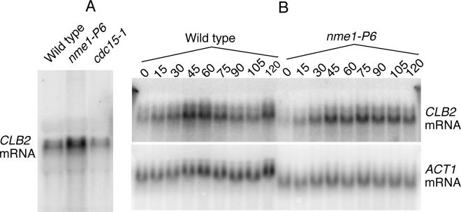
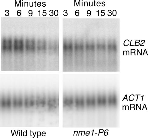
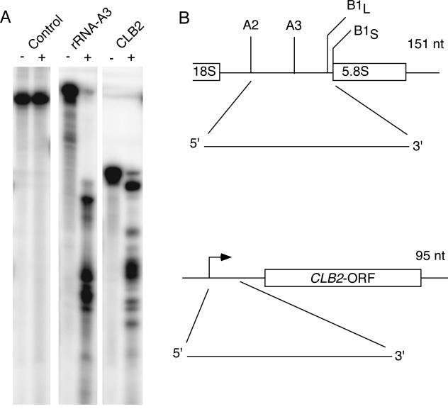
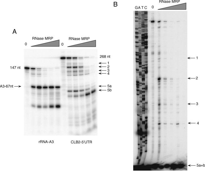
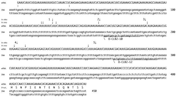
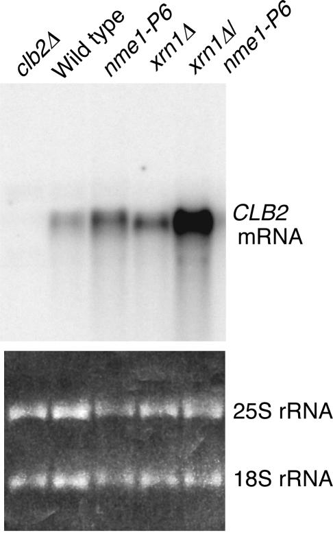
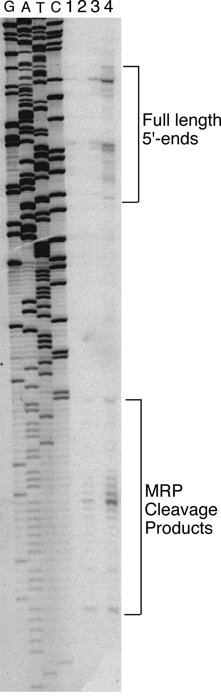
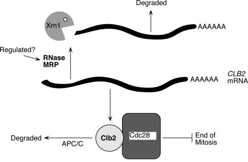
RNase mitochondrial RNA processing (RNase MRP) mutants have been shown to have an exit-from-mitosis defect that is caused by an increase in CLB2 mRNA levels, leading to increased Clb2p (B-cyclin) levels and a resulting late anaphase delay. Here we describe the molecular defect behind this delay. CLB2 mRNA normally disappears rapidly as cells complete mitosis, but the level remains high in RNase MRP mutants. This is in direct contrast to other exit-from-mitosis mutants and is the result of an increase in CLB2 mRNA stability. We found that highly purified RNase MRP cleaved the 5' untranslated region (UTR) of the CLB2 mRNA in several places in an in vitro assay. In vivo, we identified RNase MRP-dependent cleavage products on the CLB2 mRNA that closely matched in vitro products. Disposal of these products was dependent on the 5'-->3' exoribonuclease Xrn1 and not the exosome. Our results demonstrate that the endoribonuclease RNase MRP specifically cleaves the CLB2 mRNA in its 5'-UTR to allow rapid 5' to 3' degradation by the Xrn1 nuclease. Degradation of the CLB2 mRNA by the RNase MRP endonuclease provides a novel way to regulate the cell cycle that complements the protein degradation machinery. In addition, these results denote a new mechanism of mRNA degradation not seen before in the yeast Saccharomyces cerevisiae.


Figures

FIG. 1.
FIG. 1.
Analysis of CLB2 mRNA levels through the cell cycle. (A) Comparison of CLB2 mRNA levels between wild-type and mutant strains carrying the nme1-P6 or cdc15-1 mutation. Cells were grown at 24°C, shifted to 37°C for 2 h, and RNA was isolated as described in Materials and Methods. An equal amount of RNA from each strain was subjected to Northern analysis. All blots were first probed for CLB2 mRNA and subsequently for ACT1 mRNA. The locations of the relevant transcripts are shown (wild-type, MES111-140; nme1-P6, MES111-P6; cdc15-1, TLG136). (B) Analysis of CLB2 mRNA levels in cell cycle-synchronized S. cerevisiae. Wild-type (MES111-140) and nme1-P6 mutant (MES111-P6) strains were grown to 106 cells/ml at 25°C in SCD, arrested in hydroxyurea for 2 h at 25°C, and then shifted to 37°C for 1 h. Cells were then washed to remove the hydroxyurea, resuspended in fresh medium, and maintained at 37°C. Total RNA was made at 0, 15, 30, 45, 60, 75, 90, 105, and 120 min after release from hydroxyurea arrest (see Materials and Methods). RNA was harvested at the indicated times, and CLB2 and ACT1 mRNA levels were measured. Cell synchrony and release from arrest were monitored by examination of cell budding and morphology. The signals corresponding to the CLB2 and ACT1 mRNAs are shown.
FIG. 2.
FIG. 2.
Degradation of CLB2 mRNA in the nme1-P6 mutant. Wild-type (MES111-140) and nme1-P6 mutant (MES111-P6) strains were arrested in hydroxyurea for 2 h at 25°C and then shifted to 37°C for 1 h. Cells were then washed to remove the hydroxyurea, resuspended in fresh medium, and maintained at 37°C for 60 min. The transcriptional inhibitor 1,10-phenanthroline was added, and RNA was harvested at the indicated times (see Materials and Methods). The CLB2 and ACT1 mRNAs are shown.
FIG. 3.
FIG. 3.
In vitro cleavage of the CLB2 5′-UTR by RNase MRP. (A) Internally labeled RNA substrates containing the rRNA A3 cleavage site (positive control), the CLB2 5′-UTR or the 5.8S rRNA (noncleaved control RNA) were generated. Highly pure yeast RNase MRP was purified with the TAP tag protocol and used in a standard RNase MRP in vitro assay (3, 22). (B) The schematic indicates the source of the RNA substrates and the sizes of the RNAs.
FIG. 4.
FIG. 4.
In vitro cleavage of the CLB2 5′-UTR and mapping of cleavage sites. (A) 3′-end-labeled RNA substrates containing the rRNA A3 cleavage site (positive control) and a 268-nucleotide CLB2 5′-UTR were generated. Highly pure yeast RNase MRP was purified with the TAP tag protocol and used in a standard RNase MRP in vitro assay (3, 22). Increasing amounts of enzyme were used in the assay: from left to right, 0 ng, 20 ng, 40 ng, 80 ng, 200 ng, and 800 ng of protein for both substrates. (B) A second RNase MRP assay was performed exactly as in panel A but then subjected to primer extension with the primer O-CLB2-12 (34). The positions of the cleavage sites are shown in Fig. 5.
FIG. 5.
FIG. 5.
CLB2 5′-UTR. Both DNA strands are shown, with the transcribed RNA above. The major transcriptional initiation sites are at positions 6 and 39. The translational initiation site is at position 400 in this figure. Sites of both in vitro cleavage by RNase MRP and mapped in vivo ends are indicated. Numbering of cleavage sites corresponds to the positions in Fig. 4. O-CLB2-10 and O-CLB2-12 are the two major oligonucleotides used in primer extension reactions.
FIG. 6.
FIG. 6.
Both RNase MRP and Xrn1 participate in CLB2 mRNA degradation. The relevant genotypes of the yeast strains used are indicated; complete genotypes can be found in Table 1 (wild-type, MES111-140; nme1-P6, MES11-P6; xrn1Δ, TLG105; xrn1Δ nme1-P6, TLG105-P6; clb2Δ, YJA103). Cells were grown at 24°C, and equal amounts of RNA (based on both rRNA staining [bottom panel] and subsequent ACT1 mRNA analysis) were subjected to Northern analysis for the CLB2 mRNA. The CLB2 mRNA is indicated.
FIG. 7.
FIG. 7.
Identification of in vivo-generated RNase MRP products. Primer extension was performed on the same RNA used in Fig. 6 (34), with an oligonucleotide that hybridizes from positions −240 to −218 (from the CLB2 translational start). Previously identified full-length and novel xrn1Δ-specific ends are indicated (14). Lanes 1 to 4, wild-type, xrn1Δ, nme1-P6, and xrn1Δ nme1-P6, respectively. A sequencing reaction generated with the same primer on a plasmid version of the CLB2 gene is shown.
FIG. 8.
FIG. 8.
Model of the role of RNase MRP in cell cycle control. Based on our results, RNase MRP is responsible for degradation of the CLB2 mRNA. This is accomplished by processing the CLB2 mRNA in its 5′-UTR, resulting in an uncapped transcript. This uncapped transcript is then efficiently degraded by the Xrn1 5′-3′ exoribonuclease. Defects in RNase MRP increase levels of the CLB2 mRNA, producing more Clb2 protein. Sustained levels of Clb2 protein keep the cyclin-dependent kinase Cdc28 active and inhibit the end of mitosis. Genetic interactions between RNase MRP and the exit from the mitosis pathway may indicate potential regulation points of RNase MRP or a bypass involving activation of the Clb2 protein degradation pathway (4).

References

    1. Aulds, J., T. Cai, and M. E. Schmitt. 2002. RNase MRP from yeast to humans, cell cycle control and cartilage hair hypoplasia. Recent Res. Dev. Mol. Cell. Biol. 3:371-378.
    1. Beelman, C. A., and R. Parker. 1995. Degradation of mRNA in eukaryotes. Cell 81:179-183. - PubMed
    1. Cai, T., and M. E. Schmitt. 2001. Characterization of ribonuclease MRP function. Methods Enzymol. 342:135-142. - PubMed
    1. Cai, T., J. Auld, T. Gill, M. Cerio, and M. E. Schmitt. 2002. The Saccharomyces cerevisiae RNase Mitochondrial RNA Processing is critical for cell cycle progression at the end of mitosis. Genetics 161:1092-1142. - PMC - PubMed
    1. Cai, T., T. R. Reilly, M. Cerio, and M. E. Schmitt. 1999. Mutagenesis of SNM1, which encodes a protein component of the yeast RNase MRP, reveals a role for this ribonucleoprotein endoribonuclease in plasmid segregation. Mol. Cell. Biol. 19:7857-7869. - PMC - PubMed

Publication types

MeSH terms