. 2004 Feb;24(3):985-96.
doi: 10.1128/MCB.24.3.985-996.2004.

Physical and functional interactions of the Arf tumor suppressor protein with nucleophosmin/B23

Affiliations

Physical and functional interactions of the Arf tumor suppressor protein with nucleophosmin/B23

David Bertwistle et al. Mol Cell Biol. 2004 Feb.

Abstract

The Arf tumor suppressor inhibits cell cycle progression through both p53-dependent and p53-independent mechanisms, including interference with rRNA processing. Using tandem-affinity-tagged p19(Arf), we purified Arf-associated proteins from mouse NIH 3T3 fibroblasts undergoing cell cycle arrest. Tagged p19(Arf) associated with nucleolar and ribosomal proteins, including nucleophosmin/B23 (NPM), a protein thought to foster the maturation of preribosomal particles. NPM is an abundant protein, only a minor fraction of which binds to p19(Arf); however, a significant proportion of p19(Arf) associates with NPM. The interaction between p19(Arf) and NPM requires amino acid sequences at the Arf amino terminus, which are also required for Mdm2 binding, as well as the central acidic domain of NPM and an adjacent segment that regulates NPM oligomerization. The interaction between p19(Arf) and NPM occurs in primary mouse embryonic fibroblasts, including those lacking both Mdm2 and p53. In an NIH 3T3 derivative cell line (MT-Arf) engineered to conditionally express an Arf transgene, induced p19(Arf) associates with NPM and colocalizes with it in high-molecular-weight complexes (2 to 5 MDa). An NPM mutant lacking its carboxyl-terminal nucleic acid-binding domain oligomerizes with endogenous NPM, inhibits p19(Arf) from entering into 2- to 5-MDa particles, and overrides the ability of p19(Arf) to retard rRNA processing.


Figures

FIG. 1.
FIG. 1.
TAP-tagged-Arf is functional. NIH 3T3 cells were infected with MSCV-IRES-GFP retroviruses expressing Arf, TAP-tagged-Arf, or GFP alone as a negative control. Two days later, cell lysates were immunoblotted with antibodies to Arf, Mdm2, and p21Cip1 (A). Like p19Arf, although to a lesser extent, TAP-tagged Arf induced expression of Mdm2 and p21Cip1. Due to the C-terminal tag, the electrophoretic migration of TAP-tagged Arf on the denaturing gel was retarded. (B) Cells were pulse labeled with [3H]uridine for 30 min and then chased for 2.5 h. Total RNA extracted from cells was separated on a gel, transferred to a membrane, and subjected to fluororadiography. RNA in the 5-FU lane was extracted from uninfected NIH 3T3 cells which were exposed to 5-fluorouridine for 20 min prior to metabolic labeling. Expression of Arf and TAP-tagged Arf and inclusion of 5-fluorouridine in the medium all inhibited rRNA processing.
FIG. 2.
FIG. 2.
Arf associates with proteins involved in ribosome biogenesis. (A) NIH 3T3 cells were infected with a retrovirus expressing TAP-tagged Arf and lysed 48 h later. Purifications were performed with lysates from cells expressing TAP-tagged Arf (lanes 2 and 3) or from uninfected cells (lane 1). TAP-tagged Arf complexes were electrophoretically resolved on denaturing gels and visualized with silver (lanes 1 and 2, 10% of purified protein loaded) or Coomassie blue (lane 3, 80% of purified protein loaded). Coomassie blue-stained bands were sequenced by mass spectrometry. (B) NIH 3T3 cells were infected with retroviruses expressing TAP-tagged full-length Arf, Arf Δ2-14, or Arf Δ2-62 and lysed 48 h later. Purifications were performed with the same amount of protein from each of the three cell lysates. Purified complexes were resolved on denaturing gels, blotted onto a polyvinylidene difluoride membrane, stained with Sypro Ruby, and visualized with a Storm imager with the blue filter. Bands corresponding to those in panel A are indicated. (C) Lysates were either left untreated (lane 1) or treated with RNase A (lane 2) prior to affinity purification from equal quantities of total protein. Recovered proteins were separated electrophoretically and stained with Coomassie blue. The lower panel shows the results of immunoblotting performed with antibodies to ribosomal protein L7.
FIG. 3.
FIG. 3.
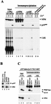
Arf forms complexes with NPM. (A, upper two panels) Lysates prepared from NIH 3T3 and MT-Arf cells treated with zinc sulfate for 24 h (+) or not (−) were precipitated with antibodies to NPM (lanes 5 to 8), p19Arf (lanes 9 to 12), or control IgG (lanes 13 and 14). Denatured immune complexes electrophoretically separated on gels were transferred to a membrane and blotted with the same antibodies. Cell lysates (10%) separated on denaturing gels were directly blotted with antibodies to NPM and p19Arf in order to estimate levels of the expressed proteins (lanes 1 to 4; see text). (A, lower panel) RNAs eluted from immune complexes were labeled with [32P]pCp and T4 RNA ligase, separated on a denaturing gel, and detected by autoradiography. (B) Primary MEFs established from day 15 embryos were propagated on a 3T3 protocol for seven passages. Cell lysates expressing both p19Arf and NPM (lane 1) were precipitated with control antibodies (lane 2) or with antibodies to NPM or Arf (lanes 3 and 4). Proteins separated on denaturing gels were immunoblotted with the same antibodies, indicated at the right of the panel. (C) Arf/Mdm2/p53 triple knockout (TKO) MEFs infected with retroviruses encoding p19Arf or the p19Arf Δ2-14 mutant were lysed and precipitated with antibodies to p19Arf (lanes 4 to 6) or nonimmune rabbit serum (NRS) (lanes 7 to 9) and subjected to immunoblotting with antibodies to Arf (top panel) or NPM (bottom panel). A portion of the cell lysate was used to demonstrate levels of protein expression (lanes 1 to 3) as in panel A.
FIG. 4.
FIG. 4.
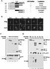
Mapping of the Arf-interacting domain in NPM. (A) Schematic representation of NPM mutants. The oligomerization domain (OligoD) (shaded), acidic domain including nuclear localization signals (NLS) (solid), heterodimerization domain (HeteroD) (stippled), and nucleic acid-binding domain (NBD) (hatched) are indicated. The results from the experiments shown in B and C are summarized on the right. (B) Localization of NPM mutants in 293T cells. Cells were transfected with pcDNA3 expression vectors containing wild-type (WT) or mutant Flag-tagged NPM variants. Exogenous proteins were detected by immunofluorescence with antibodies to the Flag epitope, and nuclei were visualized with 4′,6′-diamidino-2-phenylindole (DAPI). (C) Lysates prepared from 293T cells transiently transfected with NPM and Arf expression plasmids were separated on denaturing gels and blotted with antibodies to the Flag tag (top panel) or Arf (middle panel) to demonstrate expression of the respective proteins. Immune complexes recovered from lysates precipitated with antibodies to the Flag epitope were denatured, electrophoretically separated, and blotted with antibodies to p19Arf (bottom). (D) Transfected cells as in panel C were similarly immunoblotted to demonstrate expression of NPM variants (top panel) and p19Arf (middle panel). Immune complexes recovered from lysates precipitated with antibodies to p19Arf were blotted with antibodies to the Flag tag (bottom).
FIG. 5.
FIG. 5.
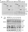
NPM modulates Arf functions. (A) NIH 3T3 and MT-Arf cells infected with the indicated retroviruses encoding NPM variants were treated (+) with zinc sulfate for 24 h or left untreated (−). Expression of NPM and p19Arf (in induced MT-Arf cells only) was confirmed by immunoblotting. The same lysates were blotted with antibodies to p53 and the p53-inducible gene product p21Cip1. Lower levels of p53 were detected in cells expressing NPM mutants C1 and C2 (lanes 20 and 22 versus 16), and this may explain the observed modest attenuation of Arf-induced proliferative arrest (Fig. 6). (B) NIH 3T3 cells (top panel) and MT-Arf cells (bottom panel) infected with the indicated retroviruses encoding NPM variants as in A were treated with zinc sulfate (+) or not (−). Newly synthesized rRNAs were analyzed by pulse-chase labeling with [3H]uridine. The indicated rRNA species separated on a gel and detected by fluoroautoradiography are indicated at the right. Note that enforced expression of C1 (lanes 5 and 6) and C2 (lanes 7 and 8) overrides the effects of Arf expression (lanes 6 and 8), whereas wild-type NPM (lane 4) and the other NPM mutants do not.
FIG. 6.
FIG. 6.
Enforced expression of NPM mutant C1 attenuates Arf-induced cell cycle arrest. NIH 3T3 and MT-Arf cells transfected with NPM and mutants C1 and C2 were pulsed for 1 h with bromodeoxyuridine (BrdU), and incorporation of bromodeoxyuridine into S-phase cells was determined by immunofluorescence. To induce p19Arf in MT-Arf cells, cultures were treated for 24 h with zinc (+) or were left untreated (−) prior to bromodeoxyuridine labeling. Incorporation into control virus-infected cells not treated with zinc was normalized to 100% in each group.
FIG. 7.
FIG. 7.
Association of endogenous and exogenous nucleophosmin and Arf. MT-Arf cells infected with retroviruses encoding GFP, full-length NPM (wild type, WT), or the C1 or C2 NPM mutant were treated with zinc sulfate for 24 h (+) or left untreated (−). Following Arf induction, cell lysates were prepared, and 10% of the total protein was separated directly on denaturing gels (A). The remainder was precipitated with the indicated antibodies (panels C to E, top) prior to separation of denatured immune complexes on gels. Control precipitations were performed with a mixture of normal mouse and rabbit IgGs (B). Separated proteins were transferred to membranes and blotted with antibodies to p19Arf (top), NPM (middle), or the Flag epitope (bottom). The monoclonal antibody to NPM used in this experiment reacts with an epitope at the C terminus of the protein and therefore recognizes both endogenously and exogenously expressed full-length NPM, but not C1 or C2. Note that Flag-tagged NPM variants form coprecipitating complexes with endogenously expressed NPM (E).
FIG. 8.
FIG. 8.
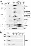
Gel filtration of complexes containing NPM and Arf. Cell lysates were prepared from MT-Arf cells infected with retroviruses encoding control protein (GFP), wild-type NPM, or the NPM C1 mutant and then treated with zinc sulfate (+) or left untreated (−). (A) Extracted cellular proteins applied and eluted from a Superose-6 column were analyzed by immunoblotting with antibodies to NPM that detect both the endogenous protein and all Flag-tagged variants. Antibodies to the Flag epitope were used to detect the C1 mutant. (B) The distribution of p19Arf recovered from induced MT-Arf cells was determined in parallel.

Similar articles

Cited by

References

    1. Bassler, J., P. Grandi, O. Gadal, T. Leβmann, E. Petfalski, D. Tollervey, J. Lechner, and E. Hurt. 2001. Identification of a 60S preribosomal particle that is closely linked to nuclear export. Mol. Cell 8:517-529. - PubMed
    1. Carnero, A., J. D. Hudson, C. M. Price, and D. H. Beach. 2000. p16INK4A and p19ARF act in overlapping pathways in cellular immortalization. Nat. Cell Biol. 2:148-155. - PubMed
    1. Colombo, E., J.-C. Marine, D. Danovi, B. Falini, and P. G. Pelicci. 2002. Nucleophosmin regulates the stability and transcriptional activity of p53. Nat. Cell Biol. 4:529-533. - PubMed
    1. Dumbar, T. S., G. A. Gentry, and M. O. Olson. 1989. Interaction of nucleolar phosphoprotein B23 with nucleic acids. Biochemistry 28:9495-9501. - PubMed
    1. Dundr, M., and M. O. Olson. 1998. Partially processed pre-rRNA is preserved in association with processing components in nucleolus-derived foci during mitosis. Mol. Biol. Cell 9:2407-2422. - PMC - PubMed

Publication types