. 2004 Feb;24(3):1096-105.
doi: 10.1128/MCB.24.3.1096-1105.2004.

Evidence for a conserved function in synapse formation reveals Phr1 as a candidate gene for respiratory failure in newborn mice

Affiliations

Evidence for a conserved function in synapse formation reveals Phr1 as a candidate gene for respiratory failure in newborn mice

Robert W Burgess et al. Mol Cell Biol. 2004 Feb.

Abstract

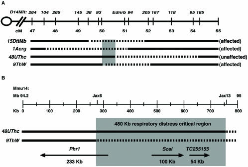
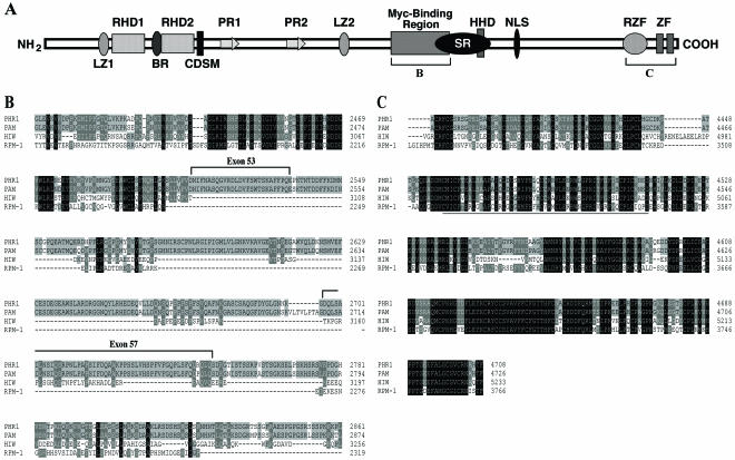
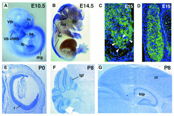
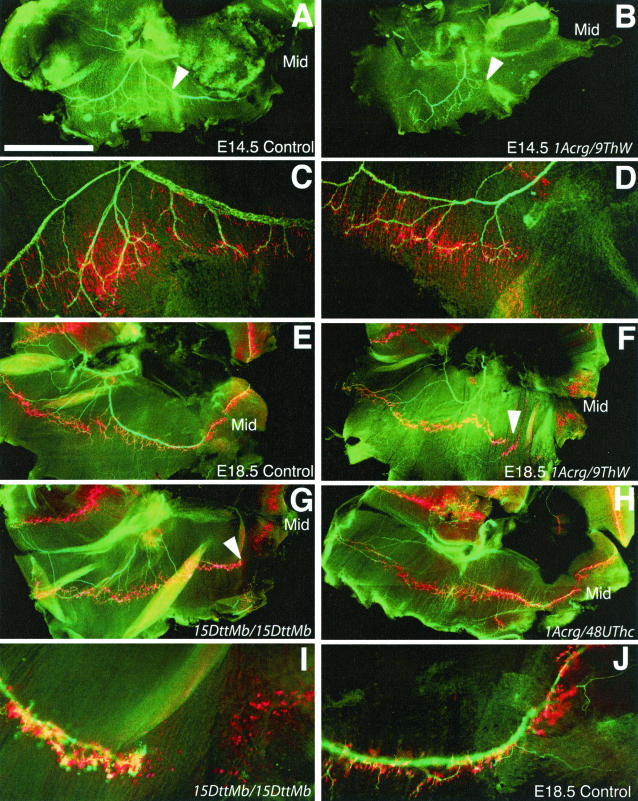
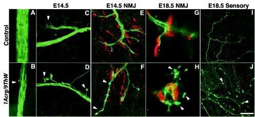
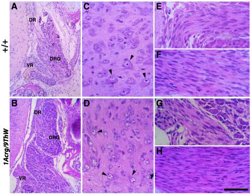
Genetic studies using a set of overlapping deletions centered at the piebald locus on distal mouse chromosome 14 have defined a genomic region associated with respiratory distress and lethality at birth. We have isolated and characterized the candidate gene Phr1 that is located within the respiratory distress critical genomic interval. Phr1 is the ortholog of the human Protein Associated with Myc as well as Drosophila highwire and Caenorhabditis elegans regulator of presynaptic morphology 1. Phr1 is expressed in the embryonic and postnatal nervous system. In mice lacking Phr1, the phrenic nerve failed to completely innervate the diaphragm. In addition, nerve terminal morphology was severely disrupted, comparable with the synaptic defects seen in the Drosophila hiw and C. elegans rpm-1 mutants. Although intercostal muscles were completely innervated, they also showed dysmorphic nerve terminals. In addition, sensory neuron terminals in the diaphragm were abnormal. The neuromuscular junctions showed excessive sprouting of nerve terminals, consistent with inadequate presynaptic stimulation of the muscle. On the basis of the abnormal neuronal morphology seen in mice, Drosophila, and C. elegans, we propose that Phr1 plays a conserved role in synaptic development and is a candidate gene for respiratory distress and ventilatory disorders that arise from defective neuronal control of breathing.


Figures

FIG. 1.
FIG. 1.
Functional map of the respiratory distress critical region. (A) Summary of complementation studies showing the original respiratory distress-defining 15DttMb deletion along with the nonrescuing 1Acrg and 9ThW deletions and the rescuing 48UThc deletion. The deleted portion of each chromosome is depicted as a dashed line, and the critical functional interval is shown in grey. Deletions are shown relative to mouse chromosome 14 from 47 to 55 cM on the Mouse Genome Database genetic map. D14Mit genetic markers used for molecular mapping studies and the piebald (Ednrb) locus are positioned along the chromosome. (B) The 480-kb respiratory distress critical region, in grey, is defined by the 48UThc and 9ThW proximal deletion breakpoints. The deletions were defined by the Jax6 and Jax13 sequence-tagged site markers. The positions and sizes of the Phr1, Scel, and TC255155 transcripts based on alignments with the genomic sequence are shown below the deletion chromosomes. Arrows indicate the direction of transcription.
FIG. 2.
FIG. 2.
Structure and conservation of PHR1. (A) Schematic representation of PHR1. The following conserved motifs were identified: LZ1 and LZ2, potential leucine zippers; RHD1 and RHD2, RCC1 homology domains; PR1 and PR2, Pam repeats; CDSM, cell division sequence motif; HHD, histone homology domain; SR, serine-rich region; NLS, nuclear localization signal; RZF, ring Zn finger domain; and ZF, potential C2H2-type zinc finger motifs. The brackets indicate the regions displayed in the protein alignments in panels B and C. (B) The Myc binding domain is conserved between PHR1 and Pam, with only the first 100 residues conserved in HIW and RPM-1. Exons 53 and 57 encode novel amino acids. (C) The C-terminal region is highly conserved between PHR1, Pam, HIW, and RPM-1. PHR1 shares 97, 65, and 55% identity across this region with Pam, HIW, and RPM-1, respectively. The ring Zn finger domain is underlined.
FIG. 3.
FIG. 3.
Phr1 is expressed in the developing nervous system. (A) Whole-mount in situ hybridization of E10.5 embryo showing Phr1 expression in peripheral neurons of the craniofacial ganglia and in the DRG flanking the spinal cord. Expression is also detected in the developing craniofacial region and forelimb bud. fn, fronto-nasal process; Vth, trigeminal ganglion; VII-VIIIth, facial and auditory ganglia; ba, first branchial arch; lb, forelimb bud. (B) Sagittal view at E14.5, showing expression throughout the mid- and hindbrain region and along the length of the developing spinal cord. md, medulla; sc, spinal cord. Transverse sections at E15.5 showed expression in cells across the dorsoventral axis of the spinal cord and in the DRG flanking the spinal cord. (C) The motor neurons of the ventral horn are robustly positive for Phr1 expression (arrowheads) (ventral is down). Motor neurons were identified based on their anatomical location and large nuclei with prominent nucleoli. (D) The sensory neuron cell bodies in the DRG are also uniformly positive for Phr1 expression. (E) Sagittal section of eye at P0, demonstrating Phr1 expression in retinal neurons. r, retina. (F) Sagittal section at P8, showing expression in the granule cell layer of the cerebellum. igl, internal granule cell layer. (G) Sagittal section at P8, showing expression in the hippocampus and throughout the cerebral cortex. ct, cerebral cortex; hip, hippocampus. In panels E, F, and G, anterior is to the right.
FIG. 4.
FIG. 4.
Innervation of the diaphragm by the phrenic nerve. (A) In control mice at E14.5, the axons of the phrenic nerve, stained green with antibodies against neurofilament and synaptophysin, have reached the diaphragm, branched, and extended both dorsally and ventrally to the full extent of the muscle. (B) In 9ThW/1Acrg littermate mice at E14.5, the phrenic nerve is less robust and fails to reach large portions of the diaphragm. In particular, the ventral portion of the muscle is not innervated. (C and D) Higher-magnification views of the regions indicated in panels A and B shows that the aneural muscle in 1Acrg/9ThW mice has a pattern of postsynaptic differentiation that is consistent with muscle that has never beeninnervated. (E) By E18.5, control mice have a similar but more elaborate pattern of motor innervation, with the nerves terminating on plaques of AChRs, stained red with rhodamine-conjugated α-bungarotoxin, on the muscle fibers. (F) In 9ThW/1Acrg mice at E18.5, the ventral diaphragm is still not innervated and the pattern of aneural muscle is similar to that at earlier ages. The defects in innervation of the diaphragm correspond to deficiency combinations that cause lethality due to respiratory distress. (G) Mice homozygous for 15DttMb show defects in the phrenic nerve that are very similar to those seen in 9ThW/1Acrg mice. (H) However, 48UThc/1Acrg mice are viable postnatally and show no defects in the diaphragm at E18.5. (I and J) Higher-magnification views of the 15DttMb diaphragm and a control diaphragm in the ventral affected region. In all cases, the right side of the diaphragm is shown, dorsal is left, ventral is right, Mid indicates the ventral midline, and the arrowheads indicate regions of incomplete innervation. Bars, 1 mm (A and B) and 1.6 mm (E to H).
FIG. 5.
FIG. 5.
Motor and sensory neuronal morphology. (A and B) The phrenic nerve contains markedly fewer axons in 9ThW/1Acrg mice than in littermate control mice. Furthermore, at E14.5, ingrowing axons show striking abnormalities in terminal morphology. (C) Control mice have tightly fascilulated axons and no visible terminal morphological defects (arrowhead). (D) 9ThW/1Acrg mice have stray axons and large varicosities at the termini (arrowheads). (E) At E14.5, neuromuscular junctions (NMJ) have formed in control mice, with nerve terminals (green) overlying AChR-rich plaques (red) on the muscle fibers. At this age, a large number of aneural receptor plaques are still evident. (F) In E14.5 9ThW/1Acrg mice, the neuromuscular junction morphology is mildly perturbed, with some overgrowth of terminals beyond the receptor plaques (lower arrowhead) and some varicose terminals (upper arrowhead). These defects are less pronounced by E15.5 (not shown) (G) In E18.5 control mice, the nerves are more robust and the receptor plaques are more condensed than at earlier stages. (H) 9ThW/1Acrg mice show similar maturation of the neuromuscular junction but also show striking sprouting of the nerve terminals beyond the receptor plaques. The terminal sprouts have varicose endings (arrowheads) similar to those seen on the ingrowing nerve at E14. (I) By E18.5, sensory neurons have also projected processes into the diaphragm from the lateral edges. These endings have a characteristic arborization pattern. (J) In 9ThW/1Acrg mice, the sensory axons are present but are of much finer caliber, have numerous varicosities, and are either more elaborately branched or less tightly fasiculated than in control mice. Bars, 36 μm (A and B), 40 μm (C to F, I, and J), and 11 μm (G and H).
FIG. 6.
FIG. 6.
Histological examination of E18.5 spinal cords. (A) The cell bodies of sensory neurons are located in the DRG. These bipolar neurons project dorsal root axons into the dorsal horn of the spinal cord. The ventral root motor axons exit the spinal cord, passing through the DRG. (B) The same anatomy is observed in 1Acrg/9ThW mutant mice. Neither pathology nor differences in cell size or number were observed in the mutants. (C) In the ventral horn of the spinal cord, motor neurons are identifiable by their large eosinophilc cytoplasm and large nuclei with prominent nucleoli (arrowheads). (D) The ventral horn of 1Acrg/9ThW mice also showed motor neurons in the proper anatomical position with normal morphology. Similarly, the axons themselves showed no pathology. Ventral roots (E and G) contain motor axons as well as elongated Schwann cell nuclei. The spinal nerves (F and H) contain both sensory afferent and motor efferent axons as well as Schwann cells. No myelination is under way at this age. DR, dorsal root; VR, ventral root. Bars, 144 μm (A and B) and 48 μm (C to H).

References

    1. Burgess, R. W., Q. T. Nguyen, Y. J. Son, J. W. Lichtman, and J. R. Sanes. 1999. Alternatively spliced isoforms of nerve- and muscle-derived agrin: their roles at the neuromuscular junction. Neuron 23:33-44. - PubMed
    1. Champliaud, M. F., H. P. Baden, M. Koch, W. Jin, R. E. Burgeson, and A. Viel. 2000. Gene characterization of sciellin (SCEL) and protein localization in vertebrate epithelia displaying barrier properties. Genomics 70:264-268. - PubMed
    1. Cole, F. S., A. Hamvas, and L. M. Nogee. 2001. Genetic disorders of neonatal respiratory function. Pediatr. Res. 50:157-162. - PubMed
    1. DeChiara, T. M., D. C. Bowen, D. M. Valenzuela, M. V. Simmons, W. T. Poueymirou, S. Thomas, E. Kinetz, D. L. Compton, E. Rojas, J. S. Park, C. Smith, P. S. DiStefano, D. J. Glass, S. J. Burden, and G. D. Yancopoulos. 1996. The receptor tyrosine kinase MuSK is required for neuromuscular junction formation in vivo. Cell 85:501-512. - PubMed
    1. DiAntonio, A., A. P. Haghighi, S. L. Portman, J. D. Lee, A. M. Amaranto, and C. S. Goodman. 2001. Ubiquitination-dependent mechanisms regulate synaptic growth and function. Nature 412:449-452. - PubMed

Publication types