. 2004 Feb;24(3):1155-67.
doi: 10.1128/MCB.24.3.1155-1167.2004.

RanBP2/Nup358 provides a major binding site for NXF1-p15 dimers at the nuclear pore complex and functions in nuclear mRNA export

Affiliations

RanBP2/Nup358 provides a major binding site for NXF1-p15 dimers at the nuclear pore complex and functions in nuclear mRNA export

Daniel Forler et al. Mol Cell Biol. 2004 Feb.

Abstract

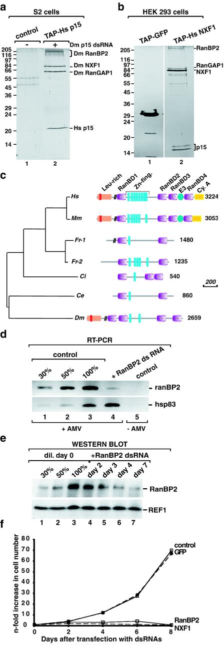
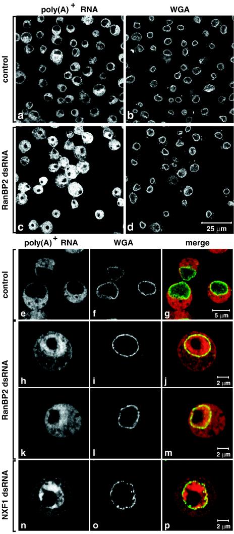
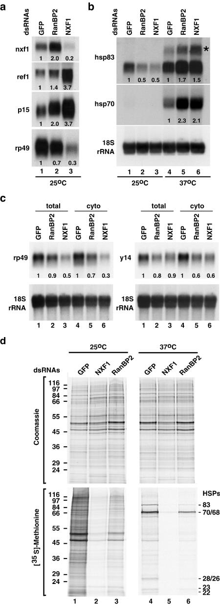
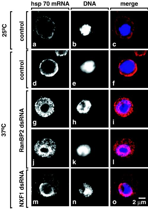
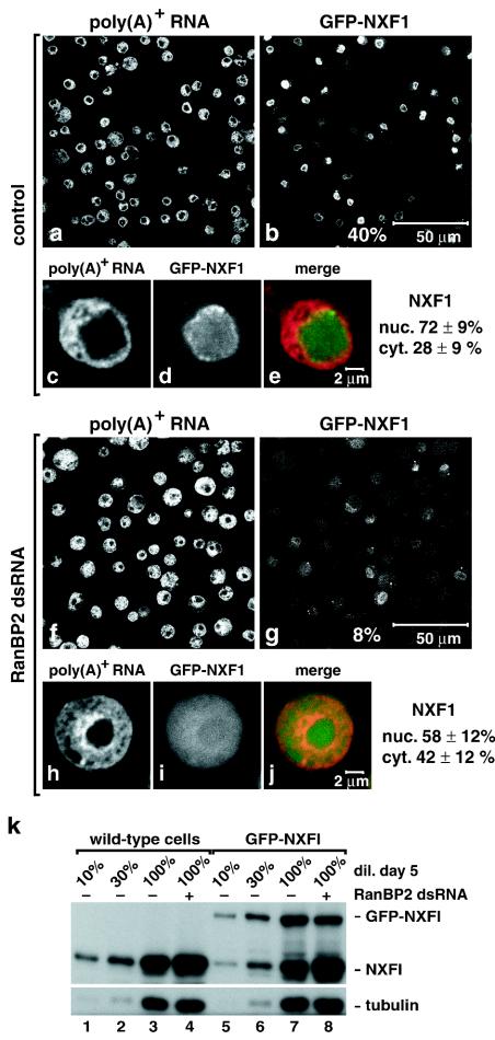
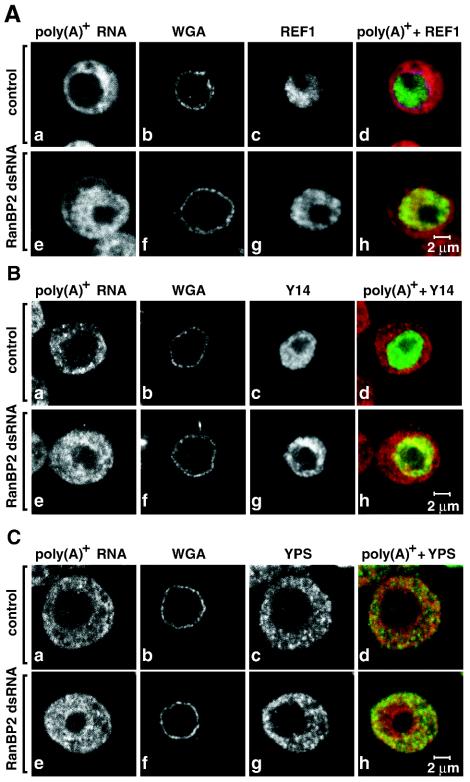
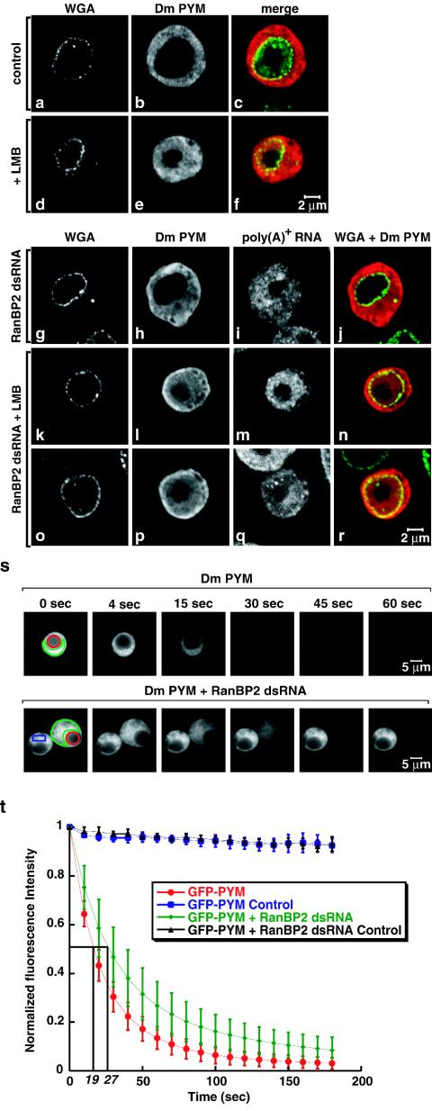
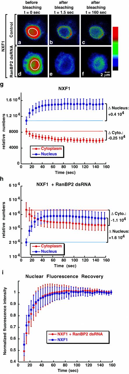
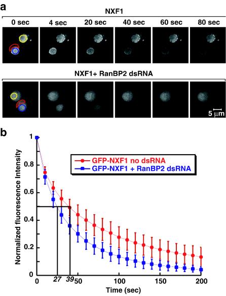
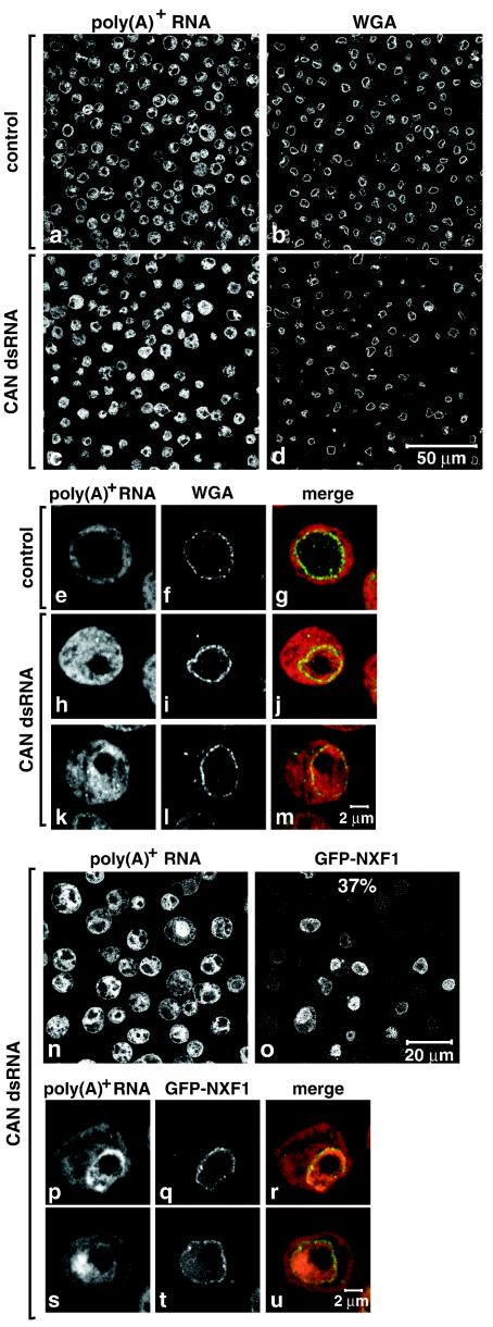
Metazoan NXF1-p15 heterodimers promote the nuclear export of bulk mRNA across nuclear pore complexes (NPCs). In vitro, NXF1-p15 forms a stable complex with the nucleoporin RanBP2/Nup358, a component of the cytoplasmic filaments of the NPC, suggesting a role for this nucleoporin in mRNA export. We show that depletion of RanBP2 from Drosophila cells inhibits proliferation and mRNA export. Concomitantly, the localization of NXF1 at the NPC is strongly reduced and a significant fraction of this normally nuclear protein is detected in the cytoplasm. Under the same conditions, the steady-state subcellular localization of other nuclear or cytoplasmic proteins and CRM1-mediated protein export are not detectably affected, indicating that the release of NXF1 into the cytoplasm and the inhibition of mRNA export are not due to a general defect in NPC function. The specific role of RanBP2 in the recruitment of NXF1 to the NPC is highlighted by the observation that depletion of CAN/Nup214 also inhibits cell proliferation and mRNA export but does not affect NXF1 localization. Our results indicate that RanBP2 provides a major binding site for NXF1 at the cytoplasmic filaments of the NPC, thereby restricting its diffusion in the cytoplasm after NPC translocation. In RanBP2-depleted cells, NXF1 diffuses freely through the cytoplasm. Consequently, the nuclear levels of the protein decrease and export of bulk mRNA is impaired.


Figures

FIG. 1.
FIG. 1.
RanBP2 forms a stable complex with NXF1-p15 dimers and is required for cell proliferation. (a) S2 cells expressing TAP-tagged hsp15 were depleted of endogenous Drosophila p15 by RNAi. Five days after addition of Drosophila p15 dsRNA, proteins bound to TAP-tagged hsp15 were purified and separated by sodium dodecyl sulfate-polyacrylamide gel electrophoresis. Following silver staining, the selected proteins were identified by mass spectrometry as described by Forler et al. (9). Lane 1, proteins purified from lysates of cells expressing the TAP tag alone; lane 2, proteins bound to TAP-tagged hp15. (b) Proteins purified from lysates of HEK 293 cells transiently expressing TAP-tagged hsNXF1. Lane 1, proteins purified from lysates of cells expressing TAP-tagged GFP; lane 2, proteins bound to TAP-tagged hsNXF1. Proteins were analyzed as described for panel a. (c) Domain organization of RanBP2 orthologs. Abbreviations: Leu-rich, leucine-rich region with a TPR homology domain (in red); RanBD, RanBP1 homology domains; Zn-fing., zinc finger motifs; E3, SUMO1 E3 ligase activity; Cy. A, cyclophilin homologous region; Ce, C. elegans; Ci, C. intestinalis; Dm, D. melanogaster; Fr, F. rubripes; Hs, Homo sapiens; Mm, Mus musculus. The vertical black bars on the schematic representation of the proteins indicate that part of the sequence is not drawn to scale. Scale bar, 200 amino acids. (d) S2 cells growing in suspension were treated with dsRNAs specific for Drosophila RanBP2, Drosophila NXF1, and GFP. Aliquots of cells were collected on day 4 and analyzed by RT-PCR. The RanBP2 mRNA levels were reduced in cells treated with RanBP2 dsRNA (lane 4), whereas the levels of the unrelated hsp83 mRNA were not affected on day 4. In lanes 1 to 3, dilutions of the cDNA isolated from untreated cells were used in the PCR to determine the efficiency of the depletion. Lane 5 shows the control sample in which the reverse transcriptase was omitted. The PCR oligonucleotides amplified a fragment of RanBP2 coding sequence corresponding to nucleotides 1100 to 1370. (e) Cells from the same experiment as in panel d were analyzed by Western blotting with monoclonal antibody mAb414. In lanes 1 to 3, dilutions of the sample isolated on day 0 were loaded to assess the efficiency of the depletion. REF1 served as a loading control. (f) Cell numbers from the same experiment as in panel d were determined every 2 days up to 8 days after addition of dsRNAs. Results are given as the increase (n-fold) in cell numbers relative to the amount used for transfection on day 0.
FIG. 2.
FIG. 2.
Depletion of endogenous RanBP2 causes the accumulation of poly(A)+ RNA within the nucleus. (a to p) S2 cells were treated for 5 days with dsRNAs corresponding to GFP (control) Drosophila RanBP2 or Drosophila NXF1. poly(A)+ RNA was detected by FISH with an oligo(dT) probe (red). The nuclear envelope was stained with Alexa 488-WGA conjugates (green).
FIG. 3.
FIG. 3.
RanBP2 acts in the mRNA export pathway. (a and b) S2 cells were treated with GFP, NXF1, and RanBP2 dsRNAs. Six days later, cells were kept at 25°C or subjected to a 1-h heat shock at 37°C. Total RNA was isolated and analyzed by Northern blot (10 μg/lane) with probes specific for the indicated mRNAs and 18S rRNA. The asterisk indicates the position of the unspliced hsp83 precursor mRNA. Numbers below the lanes represent relative mRNA expression levels normalized to 18S rRNA and set arbitrarily to 1 in control cells. (c) S2 cells were treated with GFP, NXF1, and RanBP2 dsRNAs. Total and cytoplasmic RNA were isolated 4 days (NXF1) or 5 days (GFP and RanBP2) after addition of dsRNAs. Samples were analyzed by Northern blot as described for panel a. (d) S2 cells from the same experiment shown in panels a and b were pulse labeled with [35S]methionine for 1 h. Total lysates from equivalent numbers of cells were analyzed by sodium dodecyl sulfate-polyacrylamide gel electrophoresis followed by Coomassie blue staining (upper panels) and fluorography (lower panels).
FIG. 4.
FIG. 4.
hsp70 mRNA accumulates in the nucleus of cells depleted of RanBP2. (a to o) S2 cells were treated with GFP, NXF1, and RanBP2 dsRNAs. In panels d to o, cells were shifted to 37°C for 1 h, 4 days (NXF1), or 6 days (RanBP2) after the addition of the indicated dsRNAs. hsp70 mRNA was detected by FISH. DNA was stained with Hoechst 33342. The hsp70 signal is specific, as a decrease in intensity is observed when cells were not subjected to heat stress (panels a to c). Panels show representative examples of cells. In cells depleted of RanBP2, the inhibition of hsp70 mRNA export was almost complete (panel j, 10% of the cell population) or partial with an equal distribution of the hsp70 signal between the nucleus and the cytoplasm (panel g, 80% of the cell population).
FIG. 5.
FIG. 5.
RanBP2 provides a major binding site for NXF1 at the NPC. (a to j) S2 cells constitutively expressing GFP-NXF1 (green) were treated with RanBP2 dsRNA for 5 days as indicated. poly(A)+ RNA was detected by FISH with an oligo(dT) probe (red). All panels depict optical sections, except panels d and i, which show images recorded with the pinhole of the confocal microscope open. Control panels show untreated cells. The fraction of GFP-NXF1 in the nuclear and cytoplasmic compartments of control or knockdown cells is indicated. (k) Wild-type S2 cells or cells expressing GFP-NXF1 were treated with RanBP2 dsRNA for 5 days. Total cell lysates were analyzed by Western blotting with antibodies raised to recombinant NXF1. In lanes 1 to 3 and 5 to 7, dilutions of samples isolated on day 0 were loaded to assess the expression levels of the protein. Tubulin served as a loading control. Note that in agreement with the increased levels of nxf1 mRNA (Fig. 3a) the expression levels of endogenous NXF1 were increased in the RanBP2 knockdown (lanes 4 and 8 versus lanes 3 and 7).
FIG. 6.
FIG. 6.
Depletion of RanBP2 does not alter the selectivity of the NPC. (A to C) S2 cells were fixed 5 days after the addition of RanBP2 dsRNA and labeled by FISH with an oligo(dT) probe [poly(A)+ RNA, red]. The nuclear envelope (WGA) was stained with Alexa 488-WGA conjugates. Cells shown in panel A were stained with antibodies that specifically recognize REF1 (green). Panel B shows the localization of TAP-tagged Drosophila Y14 (green). In panel C, cells were stained with antibodies raised to endogenous YPS (green). Control panels show untreated cells.
FIG. 7.
FIG. 7.
CRM1-mediated export is not inhibited in RanBP2-depleted cells. (a to f) S2 cells expressing a TAP-tagged form of Drosophila PYM were treated with LMB (Sigma) as indicated. Three hours after the addition of LMB (final concentration, 20 ng/ml), cells were fixed and the tagged protein was visualized with an anti-protein A antibody (red). Cells were double-labeled by FISH with an oligo(dT) probe [poly(A)+ RNA]. The nuclear envelope (WGA) was stained with Alexa 488-WGA conjugates (green). (g to r) S2 cells expressing a TAP-tagged Drosophila PYM were treated with RanBP2 dsRNA for 5 days. One half of the cell population was treated with LMB (k to r). Cells were labeled as described for panels a to f. (s) Control and RanBP2-depleted cells expressing GFP-PYM were selected. A nucleoplasmic region (red circle) was photobleached repetitively 130 times every 1.6 s with a 40× PlanApochromat 1.4 NA oil immersion objective. After each bleaching, the depletion of the cytoplasmic fluorescence was monitored in a confocal section (region delimited in green). (t) Quantification of cytoplasmic fluorescence loss for GFP-PYM in untreated and RanBP2-depleted cells. The loss of the cytoplasmic signal was measured and normalized to the intensity of the cytoplasmic signal before bleaching (t = 0). Curves depict mean values (± standard deviations) of the results from 8 representative cells, including those shown in panel s. Control curves show that less than 10% of the cytoplasmic fluorescence was lost from a nonbleached neighboring cell (panel s, blue rectangle) due to image acquisition between bleach pulses and the fact that the bleach pulses did not affect areas outside the bleached regions. Note that the loss of cytoplasmic fluorescence in bleached cells due to the bleach pulses cannot be evaluated. However, this loss is comparable in control and depleted cells because at steady state the fraction of GFP-PYM in the cytoplasm is similar (81% ± 2% or 81% ± 5% in 8 representative control or depleted cells, respectively).
FIG. 8.
FIG. 8.
Import of NXF1 is not significantly affected in RanBP2 depleted cells. (a to f) Control and RanBP2-depleted cells expressing GFP-NXF1 were imaged immediately before photobleaching a defined zone in the nucleus (white circle) and every 1.5 s afterwards. Colors represent relative intensities of the GFP signal (red and blue correspond to high and low intensities, respectively). (g and h) Nucleoplasmic or cytoplasmic fluorescence intensities measured over time for each individual cell were multiplied by the nuclear or cytoplasmic volumes, respectively, and the relative number of molecules in each compartment was calculated. Curves depict mean values (± standard deviations) from measurements of 8 to 9 representative cells, including those shown in panel a. The nuclear recovery (Δ Nucleus) correlates with the loss of fluorescence in the cytoplasm (Δ Cytoplasm). (i) The recovery of the nucleoplasmic signal was measured over time and normalized to the intensity of the signal when recovery was completed (160 s). Curves depict mean values (± standard deviations) from measurements of 8 to 9 representative cells, including those shown in panels a and d.
FIG. 9.
FIG. 9.
RanBP2 depletion increases the release of NXF1 in the cytoplasm. (a) Representative control and RanBP2-depleted cells expressing GFP-NXF1 were selected. A cytoplasmic region (delineated in red) was photobleached at time zero to reduce the cytoplasmic signal to background levels. Subsequently, the same region was repetitively bleached every 2.8 s. After each bleach, the depletion of the nuclear fluorescence was monitored in a confocal section (blue circle). (b) Quantitation of nuclear fluorescence loss with cytoplasmic FLIP for GFP-NXF1 in untreated and RanBP2-depleted cells. Curves depict mean values (± standard deviations) of the results from 8 representative cells, including those shown in panel a. Values are corrected for the loss of nuclear fluorescence from a nonbleached neighboring cell (panel a, yellow circle) due to image acquisition and to the bleach pulses.
FIG. 10.
FIG. 10.
Depletion of CAN inhibits mRNA export. (a to m) S2 cells were treated with a dsRNA corresponding to Drosophila CAN. poly(A)+ RNA was detected by FISH with an oligo(dT) probe (red). The nuclear envelope (WGA) was stained with Alexa 488-WGA conjugates (green). Control panels show untreated cells. (n to u) S2 cells constitutively expressing GFP-NXF1 (green) were treated with CAN dsRNA for 9 days. poly(A)+ RNA was detected by FISH (red).

References

    1. Bachi, A., I. C. Braun, J. P. Rodrigues, N. Panté, K. Ribbeck, C. von Kobbe, U. Kutay, M. Wilm, D. Görlich, M. Carmo-Fonseca, and E. Izaurralde. 2000. The C-terminal domain of TAP interacts with the nuclear pore complex and promotes export of specific CTE-bearing RNA substrates. RNA 6:136-158. - PMC - PubMed
    1. Bear, J., W. Tan, A. S. Zolotukhin, C. Tabernero, E. A. Hudson, and B. K. Felber. 1999. Identification of novel import and export signals of human TAP, the protein that binds to the CTE element of the type D retrovirus mRNAs. Mol. Cell. Biol. 19:6306-6317. - PMC - PubMed
    1. Braun, I. C., A. Herold, M. Rode, E. Conti, and E. Izaurralde. 2001. Overexpression of TAP/p15 heterodimers bypasses nuclear retention and stimulates nuclear mRNA export. J. Biol. Chem. 276:20536-20543. - PubMed
    1. Braun, I. C., A. Herold, M. Rode, and E. Izaurralde. 2002. Nuclear export of messenger RNA by TAP/NXF1 requires two nucleoporin binding sites but not p15. Mol. Cell. Biol. 15:5405-5418. - PMC - PubMed
    1. Conti, E., and E. Izaurralde. 2001. Nucleocytoplasmic transport enters the atomic age. Curr. Opin. Cell Biol. 3:310-319. - PubMed

Publication types

MeSH terms