. 2004 Feb;24(3):1206-18.
doi: 10.1128/MCB.24.3.1206-1218.2004.

ERF nuclear shuttling, a continuous monitor of Erk activity that links it to cell cycle progression

Affiliations

ERF nuclear shuttling, a continuous monitor of Erk activity that links it to cell cycle progression

Lionel Le Gallic et al. Mol Cell Biol. 2004 Feb.

Abstract

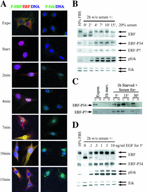
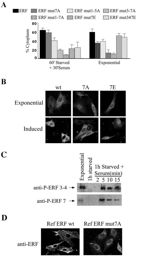
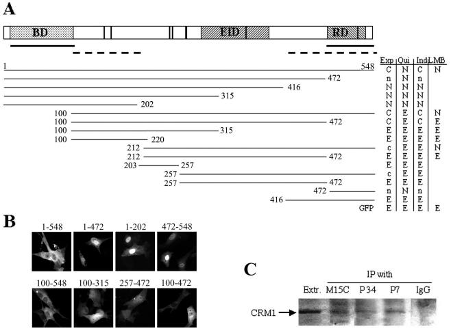
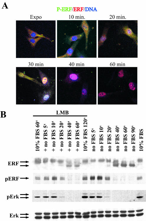
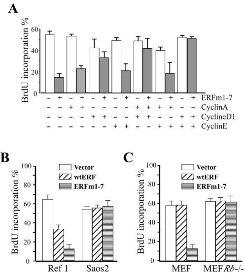
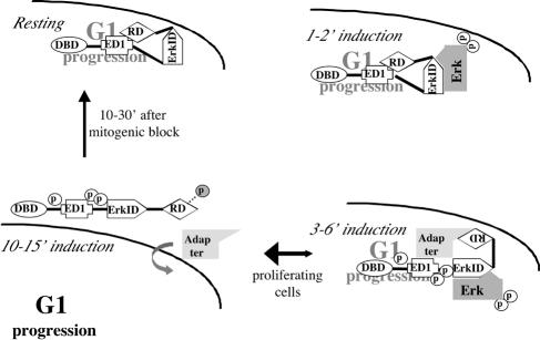
The ets domain transcriptional repressor ERF is an effector of the receptor tyrosine kinase/Ras/Erk pathway, which, it has been suggested, is regulated by subcellular localization as a result of Erk-dependent phosphorylation and is capable of suppressing cell proliferation and ras-induced tumorigenicity. Here, we analyze the effect of ERF phosphorylation on nuclear import and export, the timing of its phosphorylation and dephosphorylation in relation to its subcellular location, Erk activity, and the requirements for ERF-induced cell cycle arrest. Our findings indicate that ERF continuously shuttles between the nucleus and the cytoplasm and that both phosphorylation and dephosphorylation of ERF occur within the nucleus. While nuclear import is not affected by phosphorylation, ERF nuclear export and cytoplasmic release require multisite phosphorylation and dephosphorylation. ERF export is CRM1 dependent, although ERF does not have a detectable nuclear export signal. ERF phosphorylation and export correlate with the levels of nuclear Erk activity. The cell cycle arrest induced by nonphosphorylated ERF requires the wild-type retinoblastoma protein and can be suppressed by overexpression of cyclin. These data suggest that ERF may be a very sensitive and constant sensor of Erk activity that can affect cell cycle progression through G(1), providing another link between the Ras/Erk pathway and cellular proliferation.


Figures

FIG. 1.
FIG. 1.
ERF nuclear import is a phosphorylation-independent process. (A) Ref cells were transfected with plasmids expressing the indicated GFP-ERF fusion proteins. The localization of the fusion proteins under the indicated growth conditions was determined by measuring the GFP fluorescence under UV light. A minimum of 100 transfected cells were counted for each plasmid and growth condition, in at least three independent experiments. The mutations are as follows: 7A, T526A; 7E, T526E; 347E, S246E-S251E-T526E. (B) The localization of ERF in exponentially growing Ref-1 cells after 30 and 60 min of treatment with 300 mM LMB was determined by indirect immunofluorescence. The S17S antibody was used to determine total ERF protein (top), and the P3-4 phospho-specific antibody was used to determine the localization of ERF protein phosphorylated at S246 and S251 (middle). The localization of the activated Erks under the same conditions was determined with the phospho-specific anti-Erk mouse monoclonal antibody MAPK-YT, as a control (bottom). (C) The total amount of the phosphorylated ERF protein under the conditions described for panel B was determined by immunoblotting using the phospho-specific anti-ERF antibodies P3-4 and P7.
FIG. 2.
FIG. 2.
ERF is phosphorylated in the nucleus. (A) Ref cells were subjected to serum arrest for 2 h and consequent serum stimulation for the indicated times. The localization of the phosphorylated ERF was determined by indirect immunofluorescence using the P3-4 phospho-specific anti-ERF antibody (green), and that of total ERF was determined with the S17S anti-ERF antibody (red). Nuclei ware stained blue by TO-PRO-3 (left). At the same time points, activated Erks were detected with the phospho-specific anti-Erk monoclonal antibody MAPK-YT (green). Nuclei ware stained blue by TO-PRO-3 (right). Red and blue color colocalization shows as magenta, green and blue colocalization shows as cyan, green and red colocalization shows as yellow, and colocalization of all three shows as white. (B) Under the same conditions as in panel A, total cell extract was analyzed by immunoblotting for total and phosphorylated ERF and Erks, as indicated. The S17S, P3-4, P7, MAPK-YT, and anti-Erk specific polyclonal antibodies were used to determine total ERF, ERF phosphorylated at S246 and S251, ERF phosphorylated at T526, phosphorylated Erks, and total Erks, respectively. FBS, fetal bovine serum. (C) Ref cells were deprived of serum for 1 h and then induced with serum. At the indicated times cells were harvested and separated into nuclear and cytoplasmic fractions. The amount of the phosphorylated ERF protein in each fraction was determined by immunoblotting. Both the P3-4 (top) and the P7 (bottom) phospho-specific anti-ERF antibodies were used. (D) Ref cells were serum arrested for 2 h and then treated with the indicated amounts of epidermal growth factor for 5 min. Total cell extracts were analyzed for total and phosphorylated ERF and Erks, as for panel B.
FIG. 3.
FIG. 3.
Phosphorylation of threonine 526 plays a distinct role in nuclear export. (A) The localization of the indicated GFP-ERF fusion proteins after transfection of the corresponding plasmids into Ref-1 cells was determined as described for Fig. 1A under exponential growth and after 60 min of serum withdrawal followed by 30 min of serum stimulation. A minimum of 100 transfected cells were counted for each plasmid and growth condition in at least three independent experiments. The mutations are as follows: 7A, T526A; 1-5A, T148A-S161A-S246A-S251A-T271A; 3-7A, S246A-S251A-T271A-T357A-T526A; 1-7A, T148A-S161A-S246A-S251A-T271A-T357A-T526A; 7E, T526E; 347E, S246E-S251E-T526E. (B) Localization of the wild-type (wt) and T526A (7A) and T526E (7E) mutated forms of the ERF-GFP fusions during exponential growth and 15 min after serum induction. (C) Ref cells were subjected to serum withdrawal and stimulation. At the indicated time points the levels of the total phosphorylated ERF protein were determined by immunoblotting using the P3-4 (top) and the P7 (bottom) anti-Erf phospho-specific antibodies to determine phosphorylation at serines 246 and 251 and T526, respectively. (D) The localization of ERF was determined by indirect immunofluorescence using the S17S anti-ERF antibody in Ref-1-derived cell lines overexpressing comparable levels of wt ERF (left) or ERF carrying the T526A mutation (right).
FIG. 4.
FIG. 4.
ERF has two NLSs but no autonomous NES to mediate CRM1-dependent export. (A) Plasmids encoding GFP-ERF hybrids were transfected into Ref-1 cells, and the protein localization was determined by measuring the GFP fluorescence. The localization was determined during exponential growth 24 to 28 h after transfection (Exp column), after 3 h of serum deprivation (Qui column), after 3 h of serum deprivation and 30 min of serum stimulation (Ind column), and after 60 min of LMB treatment in the presence of serum (LMB column). A minimum of 100 transfected cells were counted for each plasmid and growth condition in at least three independent experiments, and the localization of the hybrids is indicated as follows: C, >60% cytoplasmic localization; c, 30 to 60% cytoplasmic localization; N, >60% nuclear localization; n, 30 to 60% nuclear localization; E, ubiquitous distribution. A diagram of ERF is presented at the top. BD, ets DNA-binding domain; EID, Erk interaction domain; RD, repressor domain. The vertical lines indicate the seven optimal putative Erk phosphorylation sites. Solid lines below the diagram, NLS-containing regions; dashed lines, regions contributing to nuclear export. The regions of ERF fused to the C terminus of GFP are indicated by lines below. The end points of the ERF segments are indicated by the corresponding amino acid number of the human ERF protein. (B) Representative images of the localization of GFP-ERF deletion mutations during exponential growth. (C) ERF was immunoprecipitated (IP) from cellular extracts of exponentially growing cells with the M15C, P3-4, or P7 anti-ERF specific antibodies or with control serum (IgG lane). The presence of CRM1 in the immunoprecipitated complexes and the original extract (Extr. lane) was detected by immunoblotting using an anti-CRM1 specific antibody.
FIG. 5.
FIG. 5.
ERF is dephosphorylated in the nucleus as a consequence of decreased nuclear Erk activity. (A) Ref cells were serum arrested and at the indicated time points after serum deprivation the localization of total (red) and phosphorylated (green) ERF was determined by indirect immunofluorescence as for Fig. 2A. Nuclei ware stained blue by TO-PRO-3. Red and blue color colocalization shows as magenta color, green and blue colocalization shows as cyan, green and red colocalization shows as yellow, and colocalization of all three shows as white. (B) Ref cells were serum arrested in the presence (+ no FBS lanes) or absence (no FBS lanes) of LMB. For the LMB treatment cells were grown in complete media for 1 h in the presence of LMB (10% FBS 60′ lane) and then were treated with LMB-containing serum-free media for the indicated times. Cells were also treated for 2 h with LMB in complete media (10% FBS 120′ lane) to account for any possible effects of the prolonged LMB treatment. At the indicated time points total cellular extracts were analyzed for ERF levels with the S17S antibody (top panel), phosphorylated ERF (pERF) levels with the P3-4 antibody (second panel), phosphorylated Erk1 and -2 levels with an anti-phospho-Erk antibody (third panel), and Erk1 and -2 levels with an anti-Erk antibody (fourth panel).
FIG. 6.
FIG. 6.
Nuclear ERF arrests cell proliferation in an Rb-dependent manner. (A) Ref-1 cells were cotransfected in the presence (gray bars) and absence (white bars) of ERF mut1-7A with expression plasmids encoding cyclin A, cyclin D1, and cyclin E as indicated and a plasmid encoding CD4 as a marker for the transfected cells. Twenty-four hours after transfection, cells were exposed to BrdU for 8 h, and the percentage of transfected cells that proceed to DNA replication during this 8-h window was monitored by indirect immunofluorescence with an anti-BrdU antibody. A minimum of 50 transfected cells were scored for each combination in three independent experiments. (B) Ref-1 cells and Saos2 cells were transfected with an empty vector, wild-type ERF (wtERF), or ERF mut1-7A (ERFm1-7) and a plasmid encoding CD4 to detect transfected cells. DNA synthesis was monitored as for panel A. A minimum of 50 transfected cells were scored in three independent experiments (C) Primary MEF cultures and MEF cultures from mice currying a homozygous deletion of the Rb gene (Rb−/− MEFs) were transfected with an empty vector, wtERF, or ERF mut1-7A and a plasmid encoding CD4 to detect transfected cells. DNA synthesis was monitored as for panel A. A minimum of 50 transfected cells were scored in three independent experiments.
FIG. 7.
FIG. 7.
A model for the regulation and function of ERF during Erk-mediated mitogenic stimulation. (Upper left) In the absence of Erk activity ERF is nuclear and represses the transcription of genes required for the progression through G1. (Upper right) Two to 3 min after mitogenic stimulation the activated Erk kinase translocates into the nucleus and phosphorylates ERF. (Lower right) This results in conformational changes that allow the interaction of ERF with an adaptor protein, which leads to its nuclear export. (Lower left) After the loading of ERF onto the export machinery, ERF is further and transiently phosphorylated at position T526 (grey phosphorus) in order to be released into the cytoplasm, allowing for the transcription of genes required for progression through G1. Upon the elimination of nuclear Erk activity the shuttling ERF molecules are retained in the nucleus due to their dephosphorylation (vertical arrow). In proliferating cells the export rate of ERF is high due to the Erk activity, resulting in its observed cytoplasmic localization (bidirectional horizontal arrow). The indicated domains of ERF are as follows: DBD, ets DNA-binding domain; ED1, export domain 1; ErkID, Erk interaction domain; RD, repressor domain (also contains part of export domain 2). G1 progression, transcription of genes involved in cell cycle progression through G1.

References

    1. Adachi, M., M. Fukuda, and E. Nishida. 2000. Nuclear export of MAP kinase (ERK) involves a MAP kinase kinase (MEK)-dependent active transport mechanism. J. Cell Biol. 148:849-856. - PMC - PubMed
    1. Adachi, M., M. Fukuda, and E. Nishida. 1999. Two co-existing mechanisms for nuclear import of MAP kinase: passive diffusion of a monomer and active transport of a dimer. EMBO J. 18:5347-5358. - PMC - PubMed
    1. Alt, J. R., A. B. Gladden, and J. A. Diehl. 2002. p21(Cip1) promotes cyclin D1 nuclear accumulation via direct inhibition of nuclear export. J. Biol. Chem. 277:8517-8523. - PubMed
    1. Argentini, M., N. Barboule, and B. Wasylyk. 2001. The contribution of the acidic domain of MDM2 to p53 and MDM2 stability. Oncogene 20:1267-1275. - PubMed
    1. Athanasiou, M., L. LeGallic, D. K. Watson, D. G. Blair, and G. Mavrothalassitis. 2000. Suppression of the Ewing's sarcoma phenotype by FLI1/ERF repressor hybrids. Cancer Gene Ther. 7:1188-1195. - PubMed

Publication types