. 2004 Feb;24(3):1219-31.
doi: 10.1128/MCB.24.3.1219-1231.2004.

Drosophila melanogaster MNK/Chk2 and p53 regulate multiple DNA repair and apoptotic pathways following DNA damage

Affiliations

Drosophila melanogaster MNK/Chk2 and p53 regulate multiple DNA repair and apoptotic pathways following DNA damage

Michael H Brodsky et al. Mol Cell Biol. 2004 Feb.

Abstract

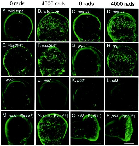
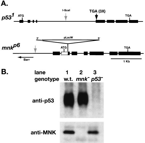
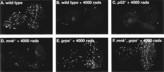
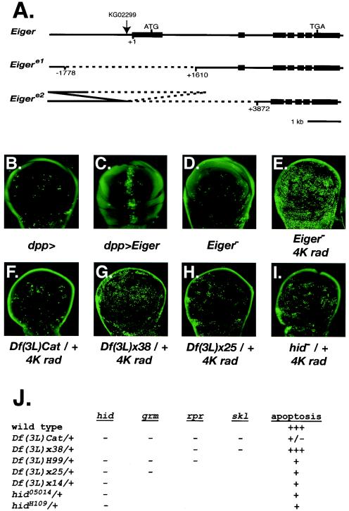
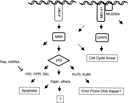
We have used genetic and microarray analysis to determine how ionizing radiation (IR) induces p53-dependent transcription and apoptosis in Drosophila melanogaster. IR induces MNK/Chk2-dependent phosphorylation of p53 without changing p53 protein levels, indicating that p53 activity can be regulated without an Mdm2-like activity. In a genome-wide analysis of IR-induced transcription in wild-type and mutant embryos, all IR-induced increases in transcript levels required both p53 and the Drosophila Chk2 homolog MNK. Proapoptotic targets of p53 include hid, reaper, sickle, and the tumor necrosis factor family member EIGER: Overexpression of Eiger is sufficient to induce apoptosis, but mutations in Eiger do not block IR-induced apoptosis. Animals heterozygous for deletions that span the reaper, sickle, and hid genes exhibited reduced IR-dependent apoptosis, indicating that this gene complex is haploinsufficient for induction of apoptosis. Among the genes in this region, hid plays a central, dosage-sensitive role in IR-induced apoptosis. p53 and MNK/Chk2 also regulate DNA repair genes, including two components of the nonhomologous end-joining repair pathway, Ku70 and Ku80. Our results indicate that MNK/Chk2-dependent modification of Drosophila p53 activates a global transcriptional response to DNA damage that induces error-prone DNA repair as well as intrinsic and extrinsic apoptosis pathways.


Figures

FIG. 1.
FIG. 1.
Drosophila p53 and mnk/Chk2 are required for IR-induced apoptosis. Imaginal wing or eye disks were dissected from untreated (A, C, E, G, I, K, M, and O) or irradiated (B, D, F, H, J, L, N, and P) third instar larvae and stained with the vital dye acridine orange. IR-induced apoptosis was observed 4 h following irradiation in wild-type (B), mei-41 (D), mus304 (F), and grps (H) mutant larvae. No induction of apoptosis was observed in mnk (J) or p53 (L) mutant larvae. Damage-induced apoptosis was restored to mnk mutant disks by a transgene containing the mnk promoter and coding sequence (M and N). Damage-induced apoptosis was restored to the posterior (shown by brackets) of p53 mutant eye disks by a transgene driving expression of a p53 cDNA under control of a Glass-responsive promoter (described in Materials and Methods).
FIG. 2.
FIG. 2.
Loss-of-function mutations in Drosophila MNK/Chk2 and p53. (A) Maps of p53 and mnk loci. Exons are depicted as rectangles, and introns are lines. Homologous recombination was used to introduce stop codons in three reading frames at the beginning of the second exon of p53, eliminating the DNA binding domain and the C-terminal tetramerization and regulatory domains. The I-SceI site used for double-strand break-mediated recombination (arrow) was present in the targeting construct but not the final mutant. The mnkp6 allele contains a P-element insertion at amino acid position 52 and an associated deletion that removes 218 nucleotides of genomic sequence, including the mnk start codon. (B) p53 and MNK protein in lysates of 2- to 16-h embryos. p53 was immunoprecipitated with mouse anti-p53 monoclonal antibody, run on an SDS-PAGE gel, and detected with guinea pig anti-p53 polyclonal antibody. Signal was detected in wild-type (lane 1) and mnk mutant (lane 2) embryos, but not in p53 mutant embryos (lane 3). These antibodies recognized a C-terminal portion of p53, indicating that translation of p53 does not reinitiate following the stop codon. MNK was detected with a polyclonal rabbit antibody (47). Signal was detected in wild-type (lane 1) and p53 mutant (lane 3) embryos, but not in mnk mutant embryos (lane 2).
FIG. 3.
FIG. 3.
Cell cycle regulation in p53 and mnk/Chk2 mutant animals. Imaginal eye disks were dissected from untreated (A) or irradiated (B to F) larvae, fixed, and stained with a phospho-specific antibody, anti-phosphorylated histone H3, which specifically recognizes mitotic cells in eukaryotes. Cell cycle arrest was assayed by the absence of mitotic cells. IR-induced arrest was observed in wild-type (A and B), p53 (I), and mnk (C and D) mutant larvae. Partial arrest was observed in grps mutant larvae (E). No arrest was observed in mnk grps double mutant larvae (F).
FIG. 4.
FIG. 4.
MNK/Chk2 is required for IR-induced modification of p53. (A) p53 protein was detected in lysates of untreated (lanes 1, 3, and 5) or irradiated (lanes 2, 4, and 6) 2- to 16-h-old embryos. p53 was immunoprecipitated with mouse anti-p53 monoclonal antibody, run on an SDS-PAGE gel, and detected with guinea pig anti-p53 polyclonal antibody. p53 mobility was reduced following irradiation of wild-type (lanes 1 and 2) but not mnk mutant (lanes 5 and 6) embryos. Phosphatase treatment (lanes 3 and 4) increased the mobility of p53 from both unirradiated (lane 3 versus lane 1) and irradiated (lane 4 versus lane 2) embryos. (B) MNK protein was detected in lysates of untreated (lanes 1, 3, and 5) or irradiated (lanes 2, 4, and 6) 2- to 16-h-old embryos. Lysates were run on an SDS-PAGE gel, and MNK was detected with rabbit anti-MNK polyclonal antibody (47). An anti-MNK signal with reduced mobility was detected in wild-type (lanes 1 and 2) and p53 mutant (lanes 5 and 6) embryos. Phosphatase treatment (lanes 3 and 4) eliminated the signal, with reduced mobility from irradiated embryos (lane 4 versus lane 2). (C to H) Adult eyes from transgenic female animals were dehydrated and viewed by scanning electron microscopy. All animals carried a transgene expressing the Gal4 transcription factor under the control of the eye-specific promoter GMR. Expression of Gal4 alone did not affect eye development and is shown here as the wild-type control. Animals were raised at either 25°C (C to F) or 18°C (G and H). (C) GMR-Gal4/+; wild-type eye morphology. (D) GMR-Gal4 GUS-p53/+; p53-dependent rough eye phenotype at 25°C. (E) GMR-Gal4 GUS-p53/GUS-GRPS; the p53-dependent rough eye phenotype was unaffected by GUS-GRPS. (F) GMR-Gal4 GUS-p53/GUS-MNK(kd); the p53-dependent rough eye phenotype was suppressed by the kinase-dead form of MNK. (G) GMR-Gal4 GUS-p53/+; p53-dependent rough eye phenotype at 18°C. (H) GMR-Gal4 GUS-p53/GUS-MNK; the p53-dependent rough eye phenotype was enhanced by wild-type MNK.
FIG. 5.
FIG. 5.
Genetic analysis of p53 targets and DNA damage-induced apoptosis. (A) Maps of Eiger mutations. Exons are depicted as rectangles, while introns are lines. The P-element insertion KG02299 (arrow) was used to generate two deletions in the Eiger locus. Eigere1 removes the first exon. Eigere2 removes the first two exons and leaves part of the KG02299 insertion. (B to F) Imaginal wing disks were dissected from third instar larvae and stained with the vital dye acridine orange to detect apoptotic cells. (B) dpp-Gal4 does not induce apoptosis. (C) dpp-Gal4 driving UAS-Eiger induces apoptosis at the anterior-posterior boundary. (D) Normal spontaneous apoptosis in homozygous Eigere1 disks. (E) Normal IR-induced apoptosis in homozygous Eigere1 disks 4 h following 4,000-rad X rays. (F to I) Imaginal wing disks were dissected from third instar larvae 4 h following 4,000-rad X rays and stained with the vital dye acridine orange. IR-induced apoptosis was reduced in Df(3L)Cat/+ (F), Df(3L)x25/+ (H), and hid05014/+ (I) and normal in Df(3L)x38/+ (G). (J) Summary of apoptotic response in animals heterozygous for deficiencies or point mutations in the 75C region. Genes disrupted in a given deficiency or mutant are indicated by a minus sign.
FIG. 6.
FIG. 6.
A working model for the organization of DNA damage response pathways in Drosophila. See the text for a discussion.

References

    1. Abdu, U., M. Brodsky, and T. Schupbach. 2002. Activation of a meiotic checkpoint during Drosophila oogenesis regulates the translation of Gurken through Chk2/Mnk. Curr. Biol. 12:1645. - PubMed
    1. Abraham, R. T. 2001. Cell cycle checkpoint signaling through the ATM and ATR kinases. Genes Dev. 15:2177-2196. - PubMed
    1. Abrams, J. M., K. White, L. I. Fessler, and H. Steller. 1993. Programmed cell death during Drosophila embryogenesis. Development 117:29-43. - PubMed
    1. Amundson, S. A., M. Bittner, P. Meltzer, J. Trent, and A. J. Fornace, Jr. 2001. Physiological function as regulation of large transcriptional programs: the cellular response to genotoxic stress. Comp. Biochem. Physiol. B 129:703-710. - PubMed
    1. Barlow, C., K. D. Brown, C. X. Deng, D. A. Tagle, and A. Wynshaw-Boris. 1997. Atm selectively regulates distinct p53-dependent cell-cycle checkpoint and apoptotic pathways. Nat. Genet. 17:453-456. - PubMed

Publication types

MeSH terms