. 2004 Feb;10(2):176-84.
doi: 10.1261/rna.5177504.

The three-dimensional architecture of the class I ligase ribozyme

Affiliations

The three-dimensional architecture of the class I ligase ribozyme

Nicholas H Bergman et al. RNA. 2004 Feb.

Abstract

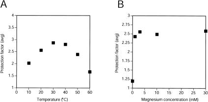
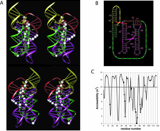
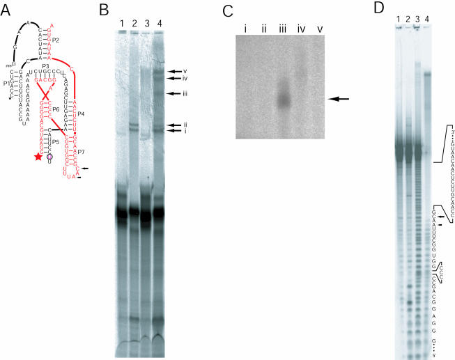
The class I ligase ribozyme catalyzes a Mg(++)-dependent RNA-ligation reaction that is chemically analogous to a single step of RNA polymerization. Indeed, this ribozyme constitutes the catalytic domain of an accurate and general RNA polymerase ribozyme. The ligation reaction is also very rapid in both single- and multiple-turnover contexts and thus is informative for the study of RNA catalysis as well as RNA self-replication. Here we report the initial characterization of the three-dimensional architecture of the ligase. When the ligase folds, several segments become protected from hydroxyl-radical cleavage, indicating that the RNA adopts a compact tertiary structure. Ribozyme folding was largely, though not completely, Mg(++) dependent, with a K(1/2[Mg]) < 1 mM, and was observed over a broad temperature range (20 degrees C -50 degrees C). The hydroxyl-radical mapping, together with comparative sequence analyses and analogy to a region within 23S ribosomal RNA, were used to generate a three-dimensional model of the ribozyme. The predictive value of the model was tested and supported by a photo-cross-linking experiment.


Figures

FIGURE 1.
FIGURE 1.
Hydroxyl-radical cleavage of the Class I ligase. (A) The class I ligase. The ligase promotes the attack of the 3′-OH of the substrate on its own 5′-α-phosphate, forming a new 3′–5′ linkage with release of pyrophosphate. Paired regions are designated P1–P7. Residues protected from hydroxyl-radical cleavage are colored red and reflect those highlighted in panel C. Residues colored blue are those for which solvent accessibility was not measured because they were too near to the ends of the RNA. (B) Gel showing hydroxyl-radical cleavage in the presence of different Mg++ concentrations. Background cleavage in the absence of iron is shown [(−)Fe], along with a lane showing cleavage under denaturing conditions (60°C, 0 mM Mg++). The gel was run for 90 min and used to collect data at positions 40–80. Nucleotides were identified by comparison to a ladder generated by partial digest of radiolabeled product using RNase T1 (not shown) and alkaline hydrolysis (OH). (C) Radioactivity profiles of lanes from the gel in panel B. Regions of substantial protection are shaded. (D) Protection factors for nt 7–115 in the presence of 10 mM Mg++ at 22°C. Protection factors are defined as the ratio of cleavage under denaturing conditions (0 mM Mg++, 60°C) to cleavage under experimental conditions. Protection factors exceeding 1.5 are labeled and colored red.
FIGURE 2.
FIGURE 2.
Temperature and Mg++ dependence of solvent accessibility in the class I ligase. (A) Protection factors were averaged for G45–C48 and G70–G75, and the average is plotted for experiments in which hydroxyl-radical cleavage was performed at varying temperatures in the presence of 10 mM Mg++. (B) Averaged protection factors are shown for the positions noted in panel A in experiments probing the ligase at different Mg++ concentrations (at 22°C).
FIGURE 3.
FIGURE 3.
A three-dimensional model for the class I ligase. (A) Two stereo views of the model. White spheres indicate those residues protected from hydroxyl-radical cleavage. Residues of particular interest are highlighted with all-atom representation. These include the residues of the proposed tandom G•A pairs and residues that both were unpaired in previous representations of the ligase secondary structure (Fig. 1A ▶) and were invariant among 25 active variants of the ligase (Ekland and Bartel 1995), all colored the same as the proximal paired regions. Also shown in all-atom representation are the four residues comprising the 2 bp flanking the ligation junction, colored according to the identity of the atoms. (B) A revised secondary structure diagram of the ligase that better reflects the arrangement of helices proposed by the tertiary model. Watson–Crick pairing is the same as in the original secondary structure (Fig. 1A ▶), except for one additional base pair, G47:C111, near the catalytic site. The color scheme reflects that of the ribbon representations in panel A. (C) Solvent accessibilities of the C4′ atoms in the modeled structure, as calculated using NACCES and a 1.4 Å sphere radius with an averaging over a window of three residues. A cut-off of 10 Å2 between accessibility and nonaccessibility is indicated by a horizontal line.
FIGURE 4.
FIGURE 4.
Crosslinking analyses of the class I ligase. (A) The secondary structure of the two-piece ribozyme used for the cross-linking study. The break in L5 facilitated insertion of a single 4SU residue (circled). The black sequence is fragment A, and the red sequence is fragment B. Stars indicate the sites of radiolabeling on the two different strands comprising the ribozyme (black 33P, red 32P). Arrows mark cross-linking sites, as mapped in panel D. (B) Separation of cross-linked ribozymes. (Lanes 1,3) The two-piece ribozyme lacks 4SU. (Lanes 2,4) The two-piece ribozyme contains 4SU. (Lanes 1,2) The RNA was not irradiated. (Lanes 3,4) Ribozymes were irradiated for 1 h before radiolabeled substrate was added. Lanes 3 and 4 contain about twice as much sample as lanes 1 and 2. The arrows with Roman numerals point to products that were excised and eluted from gel slices for subsequent relabeling. (C) Relabeling of cross-linked RNAs. The lanes marked with Roman numerals correspond to the bands in panel A. Only the RNA in lane iii was appreciably relabeled. (D) Mapping of the cross-links within an active ribozyme. (Lane 1) Unmodified, 5′-labeled fragment B RNA (red strand in A). (Lanes 2,3) Digests of labeled fragment B RNA by RNase T1, and partial alkaline hydrolysis, respectively. (Lane 4) The partial alkaline hydrolysis of the relabeled crosslinked RNA shown in panels B and C. Arrows mark the residues with the most pronounced transitions in ladder intensity, indicating the sites of the cross-links within the sequence of fragment B.

References

    1. Ban, N., Nissen, P., Hansen, J., Moore, P.B., and Steitz, T.A. 2000. The complete atomic structure of the large ribosomal subunit at 2.4 Å resolution. Science 289: 905–920. - PubMed
    1. Bartel, D.P. 1999. Re-creating an RNA replicase. In The RNA world, 2nd ed. (eds. R.F. Gesteland et al.), pp. 143–162. Cold Spring Harbor Laboratory Press, Cold Spring Harbor, NY.
    1. Bergman, N.H., Johnston, W.K., and Bartel, D.P. 2000. Kinetic framework for ligation by an efficient RNA ligase ribozyme. Biochemistry 39: 3115–3123. - PubMed
    1. Cate, J.H., Gooding, A.R., Podell, E., Zhou, K., Golden, B.L., Kundrot, C.E., Cech, T.R., and Doudna, J.A. 1996. Crystal structure of a group I ribozyme domain: Principles of RNA packing. Science 273: 1678–1685. - PubMed
    1. Celander, D.W. and Cech, T.R. 1990. Iron(II)-ethylenediaminetetraacetic acid catalyzed cleavage of RNA and DNA oligonucleotides: Similar reactivity toward single- and double-stranded forms. Biochemistry 29: 1355–1361. - PubMed

Publication types