. 2004 Feb;10(2):200-9.
doi: 10.1261/rna.5230104.

eIF4A3 is a novel component of the exon junction complex

Affiliations

eIF4A3 is a novel component of the exon junction complex

Chia C Chan et al. RNA. 2004 Feb.

Abstract

The exon junction complex (EJC) is a protein complex that assembles near exon-exon junctions of mRNAs as a result of splicing. EJC proteins play important roles in postsplicing events including mRNA export, cytoplasmic localization, and nonsense-mediated decay. Recent evidence suggests that mRNA translation is also influenced by the splicing history of the transcript. Here we identify eIF4A3, a DEAD-box RNA helicase and a member of the eIF4A family of translation initiation factors, as a novel component of the EJC. We show that eIF4A3 associates preferentially with nuclear complexes containing the EJC proteins magoh and Y14. Furthermore, eIF4A3, but not the highly related eIF4A1 or eIF4A2, preferentially associates with spliced mRNA. In vitro splicing and mapping experiments demonstrate that eIF4A3 binds mRNAs at the position of the EJC. Using monoclonal antibodies, we show that eIF4A3 is found in the nucleus whereas eIF4A1 and eIF4A2 are found in the cytoplasm. Thus, eIF4A3 likely provides a splicing-dependent influence on the translation of mRNAs.


Figures
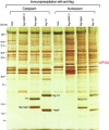
FIGURE 1.
FIGURE 1.
Identification of eIF4A3 as a flag-magoh and flag-Y14 complex associated protein in vivo by mass spectrometry. Nucleoplasmic (right) and cytoplasmic (left) extracts from parental Tet-On (mock), flag-hnRNP C1, flag-magoh, and flag-Y14 cell lines were incubated with anti-flag antibody beads and bound proteins were eluted as described in Materials and Methods. Eluted proteins were resolved by SDS-PAGE and detected by silver staining. The protein migrating with an apparent molecular weight of 47 kD was identified by mass spectrometry as eIF4A3.
FIGURE 2.
FIGURE 2.
Amino acid sequence alignment of human eIF4A translation initiation factors. Human eIF4A3 (CAA56074), eIF4A2 (BAA06336), and eIF4A1 (P04765) amino acid sequences were aligned using the ClustalW alignment program. Identical and conserved residues are indicated by dark gray and light gray shading, respectively. Peptides identified by mass spectrometry are shown in red. The conserved DEAD-box RNA helicase domain extends from amino acids ca. 82 to 372.
FIGURE 3.
FIGURE 3.
Characterization of the anti-eIF4A3 monoclonal antibody 3F1. (A) 3F1 recognizes a single band migrating at approximately 47 kD by Western blotting on total HeLa cell extract. Molecular weight markers (in kilodaltons) are shown on the left. (B) 3F1 specifically recognizes eIF4A3. Recombinant His-eIF4A3, GST-eIF4A3, GST-eIF4A2, and GST-eIF4A1 proteins were either stained with Coomassie (left panel, Protein Input) or transferred onto a nitrocellulose membrane and analyzed by Western blotting with 3F1 (right panel, 3F1 Western). (C) 3F1 immunoprecipitates eIF4A3 and not eIF4A1 or eIF4A2. Flag-eIF4A3, flag-eIF4A2, and flag-eIF4A3 were produced by in vitro transcription and translation in rabbit reticulocyte lysate in the presence of [35S]methionine and immunoprecipitated with 3F1. Bound proteins were resolved by SDS-PAGE and visualized by fluorography (right panel, 3F1 Immunoprecipitation). Input represents 10% of the amount used for immunoprecipitation (left panel, TNT Input 10%).
FIGURE 4.
FIGURE 4.
eIF4A3 localizes to the nucleoplasm by immunofluorescence. (A) Characterization of the localization of endogenous eIF4A3 in HeLa cells with anti-eIF4A3 antibody 3F1. Immunofluorescence analysis of transiently transfected myc-eIF4A3 (B), myc-eIF4A1 (C), and myc-eIF4A2 (D) with anti-myc antibody 9H10.
FIGURE 5.
FIGURE 5.
eIF4A3 associates with nuclear magoh and Y14 complexes in vivo. Nucleoplasmic (left) and cytoplasmic (right) extracts from parental Tet-On (mock), flag-hnRNP C1, flag-magoh, and flag-Y14 cell lines were incubated with anti-flag antibody beads, and bound proteins were eluted as described in Materials and Methods. Eluted proteins were resolved by SDS-PAGE and analyzed by Western blotting with anti-eIF4A3 antibody (3F1, top panel) and anti-flag antibody (M2, middle panel). Nucleoplasmic and cytoplasmic levels of eIF4A3 in total extracts are shown in the bottom panel and represent 2% of the amount used for immunoprecipitation.
FIGURE 6.
FIGURE 6.
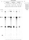
eIF4A3 binds spliced but not intronless mRNAs in vitro. (A) [32P]-labeled CDC pre-mRNA was spliced in total HeLa cell extracts. In vitro splicing reactions were immunoprecipitated with 4F4 (α-hnRNP C1), SP2/0 (control), 4C4 (α-Y14), and 3F1 (α-eIF4A3). Associated RNAs were analyzed on a 6% polyacrylamide denaturing gel, and revealed by autoradiography. (B) Same as in A except, [32P]-labeled CDC mRNA was used in this assay. Input represents 5% of total used in immunoprecipitation experiments. A schematic representation of RNAs is shown on the left.
FIGURE 7.
FIGURE 7.
eIF4A1 and eIF4A2 do not bind spliced mRNAs in vitro. (A) [32P]-labeled CDC pre-mRNA was spliced in total extracts from HeLa cells transiently transfected with flag-eIF4A3, flag-eIF4A2, or flag-eIF4A1. Splicing reactions were immunoprecipitated with SP2/0 (control), 4C4 (α-Y14), and M2 (α-flag), and coimmunoprecipitated RNAs were analyzed on a 6% polyacrylamide denaturing gel. Inputs represent 5% of total reactions used for immunoprecipitation. A schematic representation of RNAs is shown on the left. (B) Splicing extracts were resolved by SDS-PAGE and analyzed by Western blotting with the anti-flag monoclonal antibody M2.
FIGURE 8.
FIGURE 8.
eIF4A3 binds at the position of the exon junction complex. (A) [32P]-labeled Ad2 pre-mRNA was spliced in total HeLa cell extracts, incubated with A5 and A10 oligonucleotides, and subjected to RNase H digestion. Digested splicing reactions were immunoprecipitated with nonimmune antibody SP2/0 (control), H20 (α-cap), 4C4 (α-Y14), and 3F1 (α-eIF4A3). Coimmunoprecipitated RNA fragments were isolated and analyzed on 10% polyacrylamide denaturing gel. Inputs represent 5% of total reactions used for immunoprecipitation. (B) Schematic representation of the RNA fragments generated by RNase H cleavage of spliced Ad2 mRNA. The fragment associated with eIF4A3 is shown in red and the 5′ most fragment immunoprecipitated with the anti-cap antibody is shown in green.
FIGURE 9.
FIGURE 9.
eIF4A3 binds TAP and Aly/REF in vitro. The indicated proteins were produced by in vitro transcription and translation in reticulocyte lysate in the presence of [35S]methionine, and incubated with 5 μg of purified recombinant GST-eIF4A3 (middle panel) or GST-eIF4A1 (right panel) coupled to glutathione beads. Proteins bound to GST-eIF4A3 or GST-eIF4A1 were resolved by SDS-PAGE and visualized by fluorography. Input represents 2% of the in vitro translated reaction used for binding (left panel).

References

    1. Blencowe, B.J., Issner, R., Nickerson, J.A., and Sharp, P.A. 1998. A coactivator of pre-mRNA splicing. Genes & Dev. 12: 996–1009. - PMC - PubMed
    1. Bochnig, P., Reuter, R., Bringmann, P., and Lührmann, R. 1987. A monoclonal antibody against 2,2,7-trimethylguanosine that reacts with intact, class U, small nuclear ribonucleoproteins as well as with 7-methylguanosine-capped RNAs. Eur. J. Biochem. 168: 461–467. - PubMed
    1. Choi, Y.D. and Dreyfuss, G. 1984. Isolation of the heterogeneous nuclear RNA-ribonucleoprotein complex (hnRNP): A unique supramolecular assembly. Proc. Natl. Acad. Sci. 81: 7471–7475. - PMC - PubMed
    1. Dostie, J. and Dreyfuss, G. 2002. Translation is required to remove Y14 from mRNAs in the cytoplasm. Curr. Biol. 12: 1060–1067. - PubMed
    1. Dreyfuss, G., Matunis, M.J., Pinol-Roma, S., and Burd, C.G. 1993. hnRNP proteins and the biogenesis of mRNA. Annu. Rev. Biochem. 62: 289–321. - PubMed

Publication types

MeSH terms