Comparative Study
. 2004 Feb;10(2):240-53.
doi: 10.1261/rna.5153204.

The conserved RNA recognition motif 3 of U2 snRNA auxiliary factor (U2AF 65) is essential in vivo but dispensable for activity in vitro

Affiliations
Comparative Study

The conserved RNA recognition motif 3 of U2 snRNA auxiliary factor (U2AF 65) is essential in vivo but dispensable for activity in vitro

Hiren Banerjee et al. RNA. 2004 Feb.

Abstract

The general splicing factor U2AF(65) recognizes the polypyrimidine tract (Py tract) that precedes 3' splice sites and has three RNA recognition motifs (RRMs). The C-terminal RRM (RRM3), which is highly conserved, has been proposed to contribute to Py-tract binding and establish protein-protein contacts with splicing factors mBBP/SF1 and SAP155. Unexpectedly, we find that the human RRM3 domain is dispensable for U2AF(65) activity in vitro. However, it has an essential function in Schizosaccharomyces pombe distinct from binding to the Py tract or to mBBP/SF1 and SAP155. First, deletion of RRM3 from the human protein has no effect on Py-tract binding. Second, RRM123 and RRM12 select similar sequences from a random pool of RNA. Third, deletion of RRM3 has no effect on the splicing activity of U2AF(65) in vitro. However, deletion of the RRM3 domain of S. pombe U2AF(59) abolishes U2AF function in vivo. In addition, certain amino acid substitutions on the four-stranded beta-sheet surface of RRM3 compromise U2AF function in vivo without affecting binding to mBBP/SF1 or SAP155 in vitro. We propose that RRM3 has an unrecognized function that is possibly relevant for the splicing of only a subset of cellular introns. We discuss the implications of these observations on previous models of U2AF function.


Figures
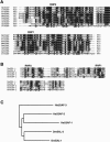
FIGURE 1.
FIGURE 1.
The RRM3 domain is highly conserved. (A) Amino acid sequence alignment of U2AF65 RRM3 from various organisms. The RRM3 sequences are from different organisms: Hs, Homo sapiens; Sp, Schizosaccharomyces pombe; Mm, Mus musculus; Sc, Saccharomyces cerevisiae; Ce, Caenorhabditis elegans; Cb, Caenorhabditis briggsiae; Dm, Drosophila melanogaster, Np, Nicotiana plumbaginifolia; Lc, Lycopersicon esculentum; At, Arabidopsis thaliana. (B) Alignment of the three RRMs of the human U2AF65 and two RRMs of the Drosophila SXL is shown. (C) Evolutionary relationship among the RRMs of U2AF65 and SXL. Alignment was done using ClustalW.
FIGURE 2.
FIGURE 2.
(Top) Deletion of RRM3 has no effect on Py-tract binding. Proteins 123 and 12 represent RRM123 and RRM12 of U2AF65, respectively. (Bottom) Schematics of RNAs, with respect to the length of Py tracts (filled boxes) and flanking sequences (open boxes). The Py tracts are: tra-NSS, tra non-sex-specific (mini and maxi); tra-FS, tra female-specific; AdML, adenoviral major late; α-TM(A), α-Tropomyosin digested with AccI; and α-TM(P), α-Tropomyosin digested with PvuII, as described (Patton et al. 1991; Singh et al. 1995; Banerjee et al. 2003). Given different binding affinities for various Py tracts, only relevant protein concentrations are shown for each Py tract. The protein concentrations for RRM123 were approximately: AdML, 1.23 ng/μL, 3.7 ng/μL, 11.1 ng/μL, 33.3 ng/μL, and 100 ng/μL; tra-NSS(mini), tra-NSS(maxi), 0.14 ng/μL, 0.41 ng/μL, 1.23 ng/μL, 3.7 ng/μL, 11.1 ng/μL; α-TM(A), and α-TM(P), 0.25 ng/μL, 0.74 ng/μL, 2.22 ng/μL, 6.66 ng/μL, 20 ng/μL; tra-FS 3.7 ng/μL, 11.1 ng/μL, 33.3 ng/μL, 100 ng/μL, and 303 ng/μL. The protein concentrations for RRM12 were approximately: AdML, 0.82 ng/μL, 2.5 ng/μL, 7.4 ng/μL, 22.2 ng/μL, and 66.6 ng/μL; tra-NSS(mini), tra-NSS(maxi), 0.09 ng/μL, 0.27 ng/μL, 0.82 ng/μL, 2.5 ng/μL, 7.4 ng/μL; α-TM(A), and α-TM(P), 0.17 ng/μL, 0.5 ng/μL, 1.5 ng/μL, 4.5 ng/μL, 13.5 ng/μL; tra-FS 2.5 ng/μL, 7.4 ng/μL, 22.2 ng/μL, 66.6 ng/μL, and 200 ng/μL.
FIGURE 3.
FIGURE 3.
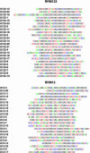
Selection–amplification of sequences by RRM123 and RRM12. Twenty-three sequences each are shown from pools 6 selected by either RRM123 (U123-series) or RRM12 (U12-series), using SELEX from a random pool of RNA.
FIGURE 4.
FIGURE 4.
RNA binding for sequences selected by either RRM123 or RRM12. The U123- and U12-series refer to the RNA sequences selected by RRM123 and RRM12, respectively. Pool 6 and three individual clones from the U123- and U12-series from pools 6 were analyzed for binding. Nucleotide sequences of these clones are given in Figure 3 ▶. −, no protein; the protein concentrations for RRM123 for each RNA panel were approximately 2.4 ng/μL, 9.6 ng/μL, 38.4 ng/μL, and 153.6 ng/μL; the protein concentrations for RRM12 for each RNA panel were approximately 1.6 ng/μL, 6.4 ng/μL, 26.6 ng/μL, and 106.4 ng/μL.
FIGURE 5.
FIGURE 5.
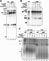
Deletion of RRM3 has no effect on the splicing of the AdML (A) or IgM (B) pre-mRNA substrates in vitro. (A) NE, nuclear extract; ΔNE, U2AF-depleted nuclear extract (Valcarcel et al. 1997). Recombinant GST-fusion proteins are shown at the top. ΔRRM3 is missing amino acids 347–475. Guanidine is the guanidine-HCl eluate from the affinity column used for U2AF depletion. The splicing product and intermediates are shown. (C) Spliceosome assembly using the full-length AdML (lanes 15) or the 3′ half of the intron (lanes 610). Splicing complexes A, B, and C, and the hnRNP complex H are indicated.
FIGURE 6.
FIGURE 6.
Schematic of the RRM3 domain of S. pombe U2AF59. (A) The amino acids that were mutagenized in different clones are underlined. For reference, amino acids for predicted β-1 to β-4 strands are also shown. A line represents amino acid identity, absence of a line represents a deletion, and one or two A residues represent alanine substitutions. The mutations are as follows: ΔRRM3a, amino acids 1–401, which correspond to amino acids 1–342 of the human U2AF65; ΔRRM3b, amino acids 1–421, in which deletion of adenine 422 generates a translation stop codon at position 425. Nomenclature for other mutants: the numbers refer to amino acid positions, the letter before the number is the wild-type amino acid, and one or more A residues following the number represent the substituted alanine(s). H, histidine; F, phenylalanine; Y, tyrosine; A, alanine. (B) A ribbon diagram of the structure of mammalian RRM3 is shown, redrawn with RasMol v2.6 using atomic coordinates of the U2AF65-RRM3/SF1 complex (Protein Data Bank, accession code 1O0P) from the NMR structure (Selenko et al. 2003). Asterisks represent positions of corresponding amino acid substitutions in the S. pombe sequence. Four β-strands (β1–β4) and three α-helices (A–C) in RRM3 are labeled. The backbone of the mBBP/SF1 peptide (amino acids 15–25) is shown.
FIGURE 7.
FIGURE 7.
The RRM3 mutants (ΔRRM3 and HFY-m) are transcribed and translated in vivo. (A) The ΔRRM3a transcript is expressed in vivo. Total cellular RNAs from either control cells (SpCR1) or cells containing the plasmid with the ΔRRM3a mutant were analyzed by RT-PCR. (Lane 1) RNA from control cells; (lanes 2 and 3) RNA from cells containing a plasmid with the ΔRRM3a mutant; (lane 3) prior to electrophoresis, the PCR product was digested with MscI and NsiI enzymes, which cut only the wild-type PCR product from the endogenous prp2 gene, shown as +RRM3, but not that from the plasmid-encoded ΔRRM3a mutant, shown as −RRM3, in which these sites have been destroyed. Asterisks in panels A and B represent primers. (B) The HFY-m transcript is expressed in vivo. Total RNA was isolated from either control cells or cells with plasmids containing either the wild-type or the HFY-m mutant genes. The 3′ end of the P4 primer perfectly base pairs with the HFY-m mutant transcript but not the wild-type transcript. In contrast, the P2 and P3 primers hybridize indistinguishably to both the wild-type and mutant transcripts, used here as internal controls. Size markers (in base pairs) are shown. (C) The RRM3 mutant proteins (ΔRRM3a and HFY-m) are stably expressed in vivo. Total cell protein lysates from either control SpCR1 cells (−) or cells containing plasmids with the wild-type (RRM123), HFY-m, or ΔRRM3a genes, indicated above respective lanes, were analyzed by Western analysis using anti-U2AF59 polyclonal antibody. Arrows indicate the U2AF59 and ΔRRM3 protein bands. (D) For loading controls, a Coommassie-stained gel of the lysates used for Western analysis in panel C is shown. Size markers (in kilodaltons) are shown.
FIGURE 8.
FIGURE 8.
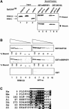
The HFY-m mutant protein interacts efficiently with splicing factors mBBP/SF1 and SAP155. (A) The RRM3 mutation HFY-m that compromises U2AF59 activity in vivo does not disrupt binding to the mammalian mBBP/SF1 or S. pombe SAP155 proteins. In vitro translated, 35S-radiolabeled proteins (wild-type RRM123, HFY-m mutant, and ΔRRM3a), indicated by arrows, were incubated with ~15–50 ng/μL of either GST-mBBP/SF1 (lanes 46) or GST-SAP155 (lanes 79), captured on GST-agarose beads without (top panel) or with (bottom panel) RNaseA, washed, and the bound fractions were analyzed on an SDS-polyacrylamide gel. (Lanes 13) A fraction (1/10) of the 35S-labeled in vitro translated proteins used for binding. (B) The binding was done as in panel A. The recombinant proteins, GST-mBBP/SF1 and GST-SAP155, were diluted threefold for each successive lane. To calculate the percent bound fraction for each lane, the signal in the GST panel served as background and that in the highest protein concentration as maximum bound (100%). (C) Phylogenetic comparison of the SF1 domain that interacts with the RRM3 domain of U2AF65 (Selenko et al. 2003). The SF1 sequences are from the following organisms: Hs, Homo sapiens; Sp, Schizosaccharomyces pombe; Mm, Mus musculus; Sc, Saccharomyces cerevisiae; Ce, Caenorhabditis elegans; Dm, Drosophila melanogaster; At, Arabidopsis thaliana. The numbers represent starting amino acids for each sequence. Alignment was done using ClustalW.
FIGURE 9.
FIGURE 9.
Model for the function of the RRM3 domain of U2AF65. The question mark (?) indicates that the nature of this interacting molecule (RNA or protein) is unknown. The broken line connecting RRM3 reflects that RRM3 is dispensable for the splicing of some introns in vitro, and likely also in vivo. RRM1 and RRM2 contact the Py tract, shown by a stretch of pyrimidine (Y) residues. The arginine-serine (RS) domain, mBBP/SF1, SAP155 and U2 snRNA interact with the branch site. For simplicity, the temporal order of interactions is not shown. mBBP/SF1 and SAP155 interact with RRM3. UAP56 and U2AF35 interact with the N-terminal region of U2AF65.

Similar articles

Cited by

References

    1. Abovich, N., Liao, X.C., and Rosbash, M. 1994. The yeast MUD2 protein: An interaction with PRP11 defines a bridge between commitment complexes and U2 snRNP addition. Genes & Dev. 8: 843–854. - PubMed
    1. Ashiya, M. and Grabowski, P.J. 1997. A neuron-specific splicing switch mediated by an array of pre-mRNA repressor sites: Evidence of a regulatory role for the polypyrimidine tract binding protein and a brain-specific PTB counterpart. RNA 3: 996–1015. - PMC - PubMed
    1. Banerjee, H., Rahn, A., Davis, W., and Singh, R. 2003. Sex lethal and U2 small nuclear ribonucleoprotein auxiliary factor (U2AF65) recognize polypyrimidine tracts using multiple modes of binding. RNA 9: 88–99. - PMC - PubMed
    1. Bashaw, G.J. and Baker, B.S. 1997. The regulation of the Drosophila msl-2 gene reveals a function for Sex-lethal in translational control. Cell 89: 789–798. - PubMed
    1. Bennett, M., Michaud, S., Kingston, J., and Reed, R. 1992. Protein components specifically associated with prespliceosome and spliceosome complexes. Genes & Dev. 6: 1986–2000. - PubMed

Publication types

MeSH terms

LinkOut - more resources