. 2004 Jan;134(1):265-74.
doi: 10.1104/pp.103.031914.

Characterization and functional identification of a novel plant 4,5-extradiol dioxygenase involved in betalain pigment biosynthesis in Portulaca grandiflora

Affiliations

Characterization and functional identification of a novel plant 4,5-extradiol dioxygenase involved in betalain pigment biosynthesis in Portulaca grandiflora

Laurent Christinet et al. Plant Physiol. 2004 Jan.

Abstract

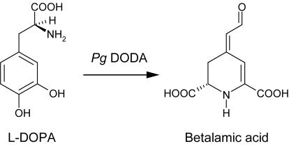
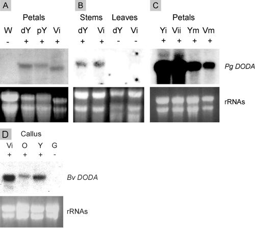
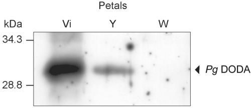
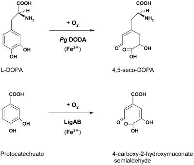
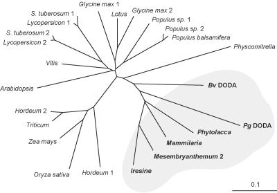
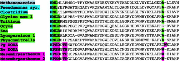
Betalains are pigments that replace anthocyanins in the majority of families of the plant order Caryophyllales. Betalamic acid is the common chromophore of betalains. The key enzyme of the betalain biosynthetic pathway is an extradiol dioxygenase that opens the cyclic ring of dihydroxy-phenylalanine (DOPA) between carbons 4 and 5, thus producing an unstable seco-DOPA that rearranges nonenzymatically to betalamic acid. A gene for a 4,5-DOPA-dioxygenase has already been isolated from the fungus Amanita muscaria, but no homolog was ever found in plants. To identify the plant gene, we constructed subtractive libraries between different colored phenotypes of isogenic lines of Portulaca grandiflora (Portulacaceae) and between different stages of flower bud formation. Using in silico analysis of differentially expressed cDNAs, we identified a candidate showing strong homology at the level of translated protein with the LigB domain present in several bacterial extradiol 4,5-dioxygenases. The gene was expressed only in colored flower petals. The function of this gene in the betalain biosynthetic pathway was confirmed by biolistic genetic complementation in white petals of P. grandiflora genotypes lacking the gene for color formation. This gene named DODA is the first characterized member of a novel family of plant dioxygenases phylogenetically distinct from Amanita sp. DOPA-dioxygenase. Homologs of DODA are present not only in betalain-producing plants but also, albeit with some changes near the catalytic site, in other angiosperms and in the bryophyte Physcomitrella patens. These homologs are part of a novel conserved plant gene family probably involved in aromatic compound metabolism.


Figures

Figure 1.
Figure 1.
Substrate and product of the Pg DODA: l-DOPA (dihydroxy-Phe) is a widespread product of plant secondary metabolism. Betalamic acid is the chromophore of betalains (betacyanins and betaxanthins) and is formed spontaneously from the 4,5-seco-DOPA intermediate resulting from the enzymatic activity (see Fig. 5).
Figure 2.
Figure 2.
RNA gel-blot analysis of DODA gene expression in P. grandiflora flowers. Presence or absence of betalain pigments (no Pg DODA) in plant tissue is indicated by + or - signs. A, Pg petals of different genotypes (W, white cc, -, -; dY, deep yellow C-, rr, ii; pY, pale yellow C-, rr, I-; Vi, violet C-, R-, ii). B, Pg stems and leaves. C, Expression at different stages of bud development (with yellow immature [Yi], yellow mature [Ym], violet immature [Vii], and violet mature [Vim]). D, RNA gel-blot analysis of expression of the putative red beet (Bv) DODA in Bv colored calli containing betalains and green calli lacking betalain (Vi, violet; O, orange; Y, yellow; and G, green).
Figure 3.
Figure 3.
Southern-blot analysis of the Pg DODA gene. Washes in SSC buffer were done to exclude the hybridization of sequences with less than 95% of homology.
Figure 4.
Figure 4.
Western-blot analysis of DODA in P. grandiflora petals of different colors (Vi, violet; Y, yellow; and W, white). Pg DODA (29.9 kD) is present in high amount in violet petals, less in yellow petals, and absent from non-pigmented white petals.
Figure 5.
Figure 5.
The l-DOPA substrate of Pg 4,5-DOPA extradiol dioxygenase DODA and the PCA substrate for the bacterial LigAB differ in the complexity of the chain containing the carboxyl group (position 1).
Figure 6.
Figure 6.
The specificity of the Pg DODA gene is demonstrated by biolistic complementation of the betalain pathway in the white petals of P. grandiflora plants deficient in DODA. A pNco DODA expression vector containing full-length Pg DODA driven by a CaMV promoter has been used with pDsRed2 vector as a positive control. A, Yellow spots revealed after biolistic transformation of a white petal from a plant with yellow genetic background. B, Close-up of a cell accumulating betaxanthins in its vacuole. C, The same cell displaying the DsRed2 fluorescent protein modified to an orange one by the fluorescence of the betaxanthins. D, Violet spots revealed in a white petal from a plant with a violet genetic background. E, Close-up of a cell accumulating betacyanins. F, The same cell displaying the DsRed2 fluorescent protein. The red fluorescence hue from DsRed2 was slightly modified by filtration through yellow betaxanthin or violet betacyanin pigments (allowing for color differences between C and F). Bars = 200 μm in A and D and 20 μm in B, C, E, and F.
Figure 7.
Figure 7.
HPLC analysis of betalain pigments extracted from violet-(A) and yellow-transformed (B) cells in the white P. grandiflora background. These pigments were identified by comparing their elution profile with elution profile of the pigments extracted from violet and deep yellow P. grandiflora petals, respectively. Arrows indicate the major peaks of the violet betanin (A) and of the yellow dopaxanthin (B). The minor 3-s shift observed between the two betanin peaks is due to a slight inaccuracy of the injection process.
Figure 8.
Figure 8.
Phylogenetic analysis of Pg DODA homologs in plants. Multiple alignments from Pg DODA homologous fragment (Met-22 to Lys-163) were done with ClustalW, and the tree was created with PHYLIP. The moss (bryophyte) P. patens corresponds to the root. Betalain-producing species in the gray surface clearly form a cluster distinct from other plants. For GenBank accession numbers, see Table I.
Figure 9.
Figure 9.
Identification of one conserved pattern specific to DODA proteins from betalain-producing plants by alignment of the Pg DODA homologous sequences from different kingdoms. The conserved catalytic amino acid His-177 is followed by the pattern P-(S,A)-(N,D)-x-T-P in all homologs of betalain-producing plants, whereas at the same place, a H-N-L pattern is conserved in all archeabacteria, and a H-N-L-R pattern is conserved in all plant homologs not producing betalains. For GenBank accession numbers, see Table I.
Figure 10.
Figure 10.
Preliminary modeling with SwissPdbViewer of the catalytic site of Pg DODA (A) and the moss P. patens DODA homolog (B) based on the three-dimensional structure 1B4U of Sphingomonas sp. LigAB. Referring to Pg DODA sequence, catalytic His-177 is conserved (blue), whereas the three following residues conserved in betalain plants Pro-178-Ser-179-Asp-180 are replaced in all non-betalain plants by Asn-178-Leu-179-Arg-180, which reduce clearly the access to the catalytic site represented by the three conserved His linking the iron cofactor (red) and the two conserved His making hydrogen bonds with the substrate (blue). Pro-178-Ser-179-Asp-180 and Thr-182-Pro-183 are conserved only in betalain plants potentially participating to the substrate recognition.

References

    1. Altschul SF, Gish W, Miller W, Myers EW, Lipman DJ (1990) Basic Local Alignment Search Tool. J Mol Biol 215: 403-410 - PubMed
    1. Ashton NW, Cove DJ, Featherstone DR (1979) Isolation and physiological analysis of mutants of the moss, Physcomitrella patens, which over-produce gametophores. Planta 144: 437-442 - PubMed
    1. Boyington JC, Gaffney BJ, Amzel LM (1993) The 3-dimensional structure of an arachidonic-acid 15-lipoxygenase. Science 260: 1482-1486 - PubMed
    1. Bradford MM (1976) Rapid and sensitive method for quantitation of microgram quantities of protein utilizing principle of protein-dye binding. Anal Biochem 72: 248-254 - PubMed
    1. Clement JS, Mabry TJ (1996) Pigment evolution in the Caryophyllales: a systematic overview. Bot Acta 109: 360-367

LinkOut - more resources