

doi: 10.1073/pnas.0305803101.
Epub 2004 Jan 19.
Increased soluble amyloid-beta peptide and memory deficits in amyloid model mice overexpressing the low-density lipoprotein receptor-related protein
Affiliations
- PMID: 14732699
- PMCID: PMC327153
- DOI: 10.1073/pnas.0305803101
Item in Clipboard
Increased soluble amyloid-beta peptide and memory deficits in amyloid model mice overexpressing the low-density lipoprotein receptor-related protein
Proc Natl Acad Sci U S A.
.
Abstract
Amyloid-beta peptide (Abeta) is central to the pathogenesis of Alzheimer's disease, and the low-density lipoprotein receptor-related protein (LRP) has been shown to alter Abeta metabolism in vitro. Here, we show that overexpression of a functional LRP minireceptor in the brain of PDAPP mice results in age-dependent increase of soluble brain Abeta, with no changes in Abeta plaque burden. Importantly, soluble brain Abeta was found to be primarily in the form of monomers/dimers and to be highly correlated with deficits in spatial learning and memory. These results provide in vivo evidence that LRP may contribute to memory deficits typical of Alzheimer's disease by modulating the pool of small soluble forms of Abeta.
Figures
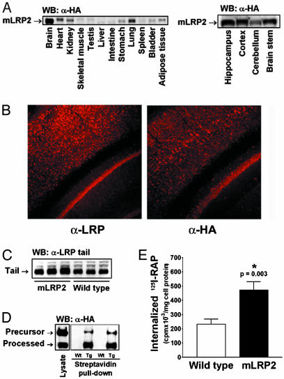
Expression of PrP-mLRP2 transgene. (A) Expression of mLRP2 was highest in the brain, and, within the brain, mLRP2 was expressed in multiple regions. (B) mLRP2 staining pattern (Right) was similar to that of endogenous LRP (Left). (C) The overall LRP expression in the brain of mLRP2 TG mice was 3.7-fold higher than in wild-type mice as detected by an antibody to the tail region of LRP, which is identical between endogenous LRP and mLRP2. (D) mLRP2 was effectively processed to the cell surface as shown by cell-surface biotinylation. (E) mLRP2 overexpression increased receptor-mediated 125I-RAP internalization by primary neuronal cultures.

Overexpression of mLRP2 in PDAPP mice increased soluble Aβ in an age-dependent manner. (A) Carbonate-soluble Aβ levels were increased in both hippocampus and cortex of aged PDAPP/LRP+ mice compared to littermate PDAPP/LRP– controls. (B) Insoluble Aβ level was also increased in the cortex but not in the hippocampus of aged PDAPP/LRP+ mice. Neither the percentage of hippocampal area covered by Aβ plaques (C) nor that of thioflavine S-positive plaques (D) differed between PDAPP/LRP+ and PDAPP/LRP– mice. *, Statistically significant differences by ANOVA.

Aβ monomers and dimers were increased in carbonate-soluble brain extracts of PDAPP/LRP+ mice. (A) By gel-filtration chromatography, most of the carbonate-soluble Aβ eluted at dimer size (≈9 kDa). Graphs shown are representative of three separate experiments in which extracts from five to six animals of each group were combined for analysis. (B) When extracts from frontal cortex of PDAPP/LRP– were separated by Tris-tricine gel, carbonate-soluble Aβ (lane 2) was detected mostly as monomers (arrow) as well as dimers (arrowhead). Aβ standards are shown in lane 1. (C) Soluble Aβ40 and Aβ42 differences between PDAPP/LRP+ and PDAPP/LRP– brain extracts before gel filtration were similar to the ones observed in the ≈9-kDa peaks shown in A.
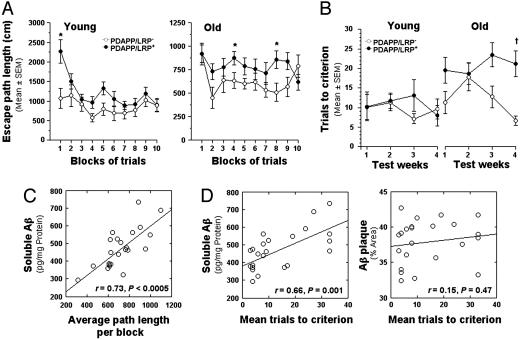
Deficits in spatial learning and memory were increased in PDAPP/LRP+ mice and correlated with soluble Aβ levels measured in the hippocampus. (A) Performance of PDAPP/LRP+ mice during standard place trials in terms of path length was inferior to that of PDAPP/LRP– mice in both young and old groups. (B) The average number of trials to reach the acquisition criterion (three consecutive trials with an average escape latency of <20 s) in both young and old groups of mice as a function of 4 weeks of training. (C) In aged mice, performance deficits observed during standard place training were highly correlated with soluble Aβ levels in the hippocampus. (D) A significant correlation was also found between mean trials-to-criterion scores in old mice during week 4 and hippocampal soluble Aβ levels but not with Aβ plaque burden. In all figures, * indicates P < 0.05, and † indicates P < 0.005 (by ANOVA).
References
-
- Hardy, J. & Selkoe, D. J. (2002) Science 297, 353–356. - PubMed
-
- Hardy, J. A. & Higgins, G. A. (1992) Science 256, 184–185. - PubMed
-
- Lendon, C. L., Talbot, C. J., Craddock, N. J., Han, S. W., Wragg, M., Morris, J. C. & Goate, A. M. (1997) Neurosci. Lett. 222, 187–190. - PubMed
-
- Wavrant-DeVrieze, F., Lambert, J. C., Stas, L., Crook, R., Cottel, D., Pasquier, F., Frigard, B., Lambrechts, M., Thiry, E., Amouyel, P., et al. (1999) Hum. Genet. 104, 432–434. - PubMed
-
- Narita, M., Holtzman, D. M., Schwartz, A. L. & Bu, G. (1997) J. Neurochem. 69, 1904–1911. - PubMed
Publication types
MeSH terms
Substances
Grants and funding
LinkOut - more resources
Full Text Sources
Other Literature Sources
Medical
Molecular Biology Databases
Miscellaneous
