. 2004 Jan 19;199(2):173-83.
doi: 10.1084/jem.20030850.

Activation of arterial wall dendritic cells and breakdown of self-tolerance in giant cell arteritis

Affiliations

Activation of arterial wall dendritic cells and breakdown of self-tolerance in giant cell arteritis

Wei Ma-Krupa et al. J Exp Med. .

Abstract

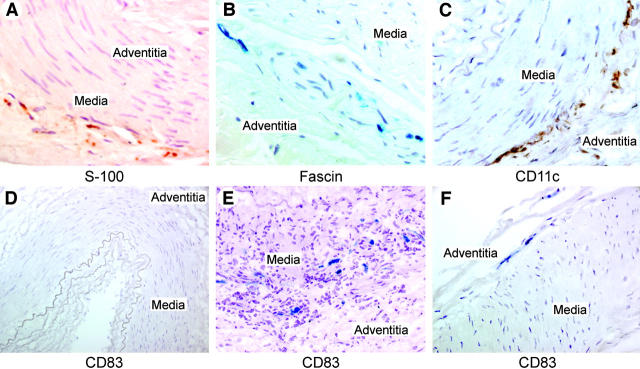
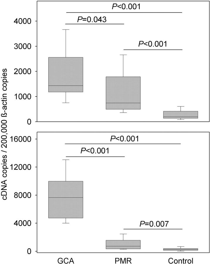
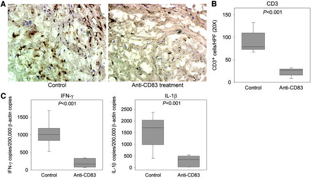
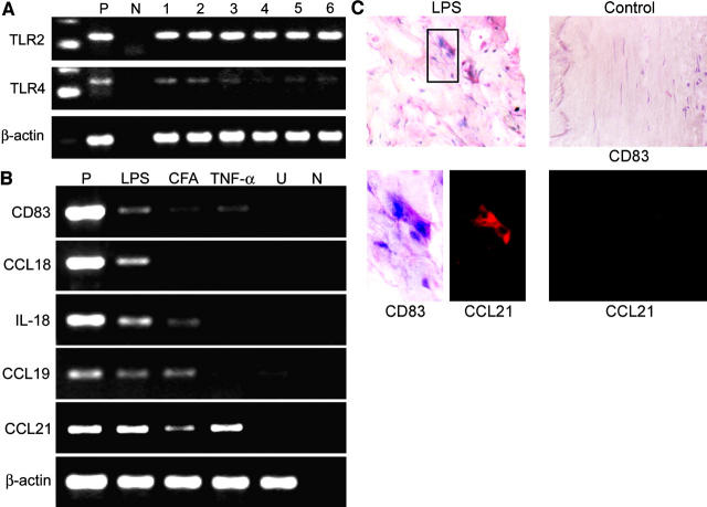
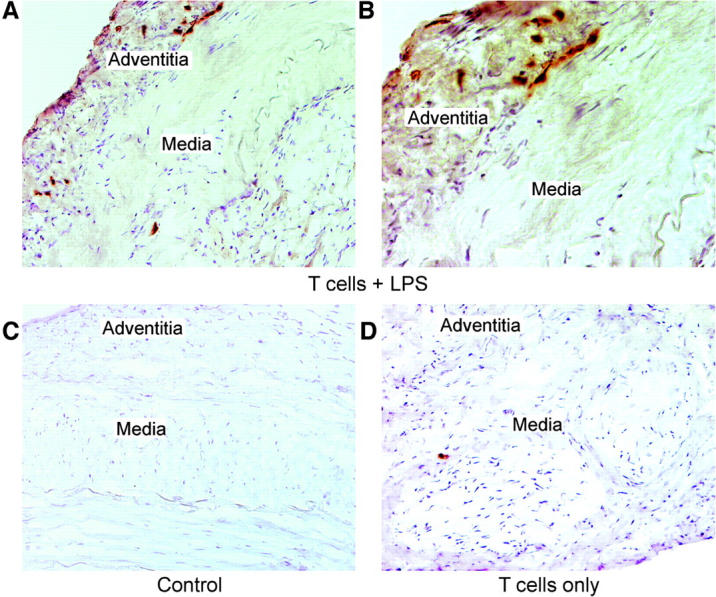
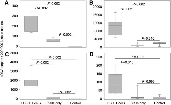
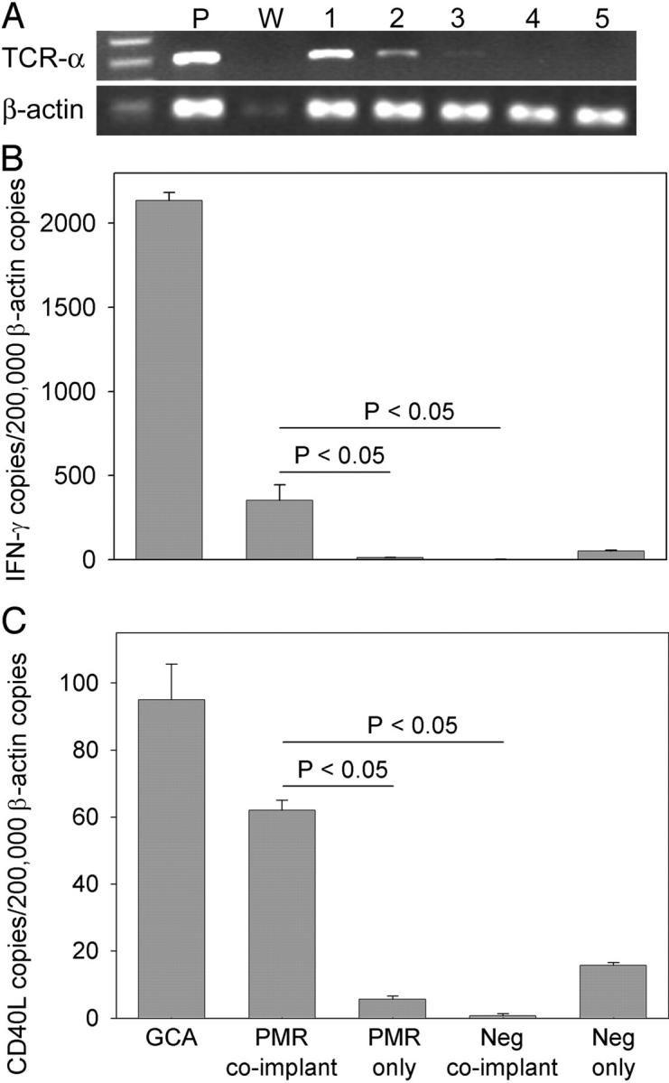
Giant cell arteritis (GCA) is a granulomatous and occlusive vasculitis that causes blindness, stroke, and aortic aneurysm. CD4(+) T cells are selectively activated in the adventitia of affected arteries. In human GCA artery-severe combined immunodeficiency (SCID) mouse chimeras, depletion of CD83(+) dendritic cells (DCs) abrogated vasculitis, suggesting that DCs are critical antigen-presenting cells in GCA. Healthy medium-size arteries possessed an indigenous population of DCs at the adventitia-media border. Adoptive T cell transfer into temporal artery-SCID mouse chimeras demonstrated that DCs in healthy arteries were functionally immature, but gained T cell stimulatory capacity after injection of lipopolysaccharide. In patients with polymyalgia rheumatica (PMR), a subclinical variant of GCA, adventitial DCs were mature and produced the chemokines CCL19 and CCL21, but vasculitic infiltrates were lacking. Human histocompatibility leukocyte antigen class II-matched healthy arteries, PMR arteries, and GCA arteries were coimplanted into SCID mice. Immature DCs in healthy arteries failed to stimulate T cells, but DCs in PMR arteries could attract, retain, and activate T cells that originated from the GCA lesions. We propose that in situ maturation of DCs in the adventitia is an early event in the pathogenesis of GCA. Activation of adventitial DCs initiates and maintains T cell responses in the artery and breaks tissue tolerance in the perivascular space.


Figures

Figure 1.
Figure 1.
DCs in normal and inflamed temporal arteries. Temporal arteries were collected from diagnostic biopsies. Patients with granulomatous inflammatory infiltrates were categorized as having GCA. Patients were diagnosed with PMR if they had a typical clinical presentation but no vascular infiltrates. Normal temporal arteries were from patients who had neither PMR nor GCA. Paraffin-embedded arteries were stained with anti–S-100 (A), antifascin (B), or anti-CD11c Ab (C) to identify DCs. Frozen sections were stained with anti-CD83 Ab to assess DC activation (D–F). Normal temporal arteries contain a population of S-100+ (red), fascin+ (blue), and CD11c+ (brown) DCs positioned at the adventitia–media border (A–C). In noninflamed arteries, these DCs lack CD83 (D). Numerous CD83+ DCs (blue) participate in the granulomatous lesions in GCA (E). In arteries from patients with PMR, adventitial DCs have acquired the activation marker CD83 (F). Original magnification, 200 (D–F) and 400 (A–C).
Figure 2.
Figure 2.
Tissue production of CCL19 and CCL21 in temporal arteries from patients with GCA or PMR and in normal arteries. Temporal arteries were collected, and cDNA was generated. Transcripts for β-actin, CCL19 (top), and CCL21 (bottom) were determined by quantitative PCR. cDNA concentrations were adjusted to 2 × 10⁵ β-actin copies. The number of chemokine transcripts is presented as box plots, with the median and 25th and 75th percentiles as the box, and the whiskers signify the 10th and 90th percentiles. Normal arteries contained minimal copies of CCL19 and CCL21 mRNA. Abundant transcripts for both chemokines were present in arteries with GCA. Arteries from patients with PMR, despite the lack of inflammatory infiltrates, contained intermediate numbers of CCL19 and CCL21 mRNA transcripts.
Figure 3.
Figure 3.
Therapeutic effects of depleting CD83+ DCs in GCA. Temporal artery–SCID mouse chimeras were generated by implanting SCID mice with segments from a GCA-affected temporal artery. The chimeras were injected on days 9–11 with anti-CD83 Ab or control Ig, and arterial grafts were harvested 1 wk later. Frozen tissue sections were immunostained with anti-CD3 Ab (A). The density of arterial wall T cell infiltrates was determined by counting CD3+ T cells (brown) on cross sections of the arteries (B). cDNA was generated from the explanted temporal arteries to quantify transcripts for β-actin, IFN-γ, and IL-1β by quantitative PCR. All cDNA concentrations were adjusted to 2 × 10⁵ β-actin copies. Copy numbers of IFN-γ– and IL-1β–specific sequences are shown as box plots as described in Fig. 2. Results are from five experiments with tissues from five different patients. Treatment with anti-CD83 Ab induced massive loss of tissue-infiltrating T cells. In parallel, IFN-γ mRNA transcripts decreased to <20% of baseline, and IL-1β mRNA transcripts decreased by >75% (C). Original magnification, 400.
Figure 4.
Figure 4.
Functional characteristics of arterial wall DCs in normal arteries. Temporal arteries were collected from patients with neither GCA nor PMR. Tissue extracts from fresh shock-frozen samples were analyzed for TLR2- and TLR4-specific sequences by PCR. All negative arteries (marked as 1–6) contained mRNA transcripts for TRL2 and TLR4 (A). To test the responsiveness of arterial DCs to triggering with blood-born TNF-α or TLR ligands, we implanted pieces of arteries into SCID mice. 7–10 d after implantation, the SCID mouse chimeras were injected with 2 μg i.v. TNF-α, 10 μg i.v. LPS, or 100 μl i.p. CFA, and the arterial grafts were harvested 48 h later. Tissue extracts from the explanted grafts were analyzed for the mRNA transcripts of β-actin, CD83, IL-18, and the chemokines CCL18, CCL19, and CCL21. After stimulation with blood-born triggers, arterial wall DCs expressed CD83+ and began to produce an array of chemokines. The effect of TNF-α was limited to the induction of CCL21, whereas LPS induced the full spectrum of chemokines. One experiment representative of three is shown (B). Immunohistochemistry confirmed that arterial DCs from LPS-treated (left), but not control (right), arteries expressed CD83 (blue) and produced CCL21 (red) (C). Original magnification, 200 (except LPS-treated CD83 and CCL21 images, which were 600×). P, positive PCR control; N, untreated mouse control; and W, negative PCR control.
Figure 5.
Figure 5.
Activated arterial DCs are capable of stimulating T cells. Temporal artery–SCID mouse chimeras were generated by implanting normal temporal arteries from HLA-DRB1*0401+ donors. 6 d after implantation, the chimeras received 10 μg LPS or PBS i.v., and 30 h later, 5 × 10⁶ alloreactive human T cell clones specific for HLA-DRB1*0401 or T cell lines derived from arterial tissue of patients with GCA were adoptively transferred into the mice. Arterial grafts were explanted and embedded in OCT. To locate human T cells in the arterial grafts, frozen tissue sections were immunostained with anti-CD3 Ab. One experiment representative of five is shown. CD3+ T cells (brown) were found in the arteries explanted from the chimeras that had been treated with LPS and had received the T cells. Tissue-infiltrating T cells accumulated along the adventitia–media junction (A and B). In the absence of LPS pretreatment, human T cells were rarely detected in the arteries (D). Grafts from animals that had received neither LPS nor T cells were free of human T cells (C). Original magnification, 200 (A, C, and D) and 400 (B).
Figure 6.
Figure 6.
Stimulation of adoptively transferred T cells by activated arterial DCs. Temporal artery–SCID mouse chimeras carrying HLA-DRB1*0401+ normal arteries were treated with LPS and adoptively transferred with alloreactive human T cells as described in Fig. 5. Explanted arterial grafts were shock frozen and used for the generation of cDNA. cDNA concentrations were adjusted to 2 × 10⁵ β-actin transcripts. Tissue concentrations of β-actin, CD83 (A), IL-18 (B), IFN-γ (C), and CD40L (D) mRNA transcripts were determined by quantitative PCR. Pretreatment with LPS induced prompt activation of DCs as indicated by the up-regulation of CD83 and IL-18 mRNA transcripts. IFN-γ–and CD40L-specific mRNA transcripts were clearly induced in arteries that had been pretreated with LPS and that received human alloreactive T cell clones. In the absence of LPS stimulation, adoptively transferred T cells induced a minimal up-regulation of CD83, but they failed to undergo in situ activation.
Figure 7.
Figure 7.
DC activation in PMR arteries is sufficient to activate disease-relevant T cells from GCA arteries. Donors of normal arteries and patients with PMR or GCA were typed for HLA-DR, and HLA-DRB1*0401+ arteries were selected. Arteries from patients with each diagnosis were implanted individually into SCID mice or three arteries (one from each disease category) were implanted into nonadjacent sites of the same mouse. After 7 d, the grafts were harvested, shock frozen, and used for the generation of cDNA. cDNA concentrations were adjusted to equal numbers of β-actin copies. One experiment representative of three is shown. Tissue-infiltrating T cells were detected by PCR analysis for TCR-α sequences (A). T cell activation in the tissues was assessed by quantifying mRNA transcripts for IFN-γ (B) and CD40L (C) by quantitative PCR. GCA arteries contained abundant TCR-α mRNA (lane 1). No T cells were detected in negative arteries (lanes 4 and 5), which remained free of TCR-α sequences even after coimplantation with inflamed arteries (lane 4). PMR arteries had a minimal signal for TCR-α if implanted individually (lane 3), but they accumulated TCR-α copies when combined with inflamed arteries (lane 2). No IFN-γ or CD40L could be detected in negative arteries. PMR arteries remained negative for IFN-γ and CD40L as long as they were isolated from arteries with typical vasculitic lesions. Once GCA and PMR arteries were combined in the same chimera, T cells from the vasculitic infiltrates migrated into the PMR arteries, where they produced IFN-γ and CD40L (B and C). Copy numbers of IFN-γ and CD40L are shown as mean ± SD for one representative experiment. P, positive PCR control; W, negative PCR control.

Similar articles

Cited by

References

    1. Weyand, C.M., and J.J. Goronzy. 2003. Vasculitis of medium- and large-size arteries. N. Engl. J. Med. 349:160–169. - PubMed
    1. Weyand, C.M. 2000. The Dunlop-Dottridge lecture: the pathogenesis of giant cell arteritis. J. Rheumatol. 27:517–522. - PubMed
    1. Hunder, G.G., and R.M. Valente. 2002. Giant cell arteritis: clinical aspects. Inflammatory Diseases of Blood Vessels. G.S. Hoffman and C.M. Weyand, editors. Marcel Dekker, New York. 425–441.
    1. Björnsson, J. 2002. Histopathology of primary vasculitic disorders. Inflammatory Diseases of Blood Vessels. G.S. Hoffman and C.M. Weyand, editors. Marcel Dekker, New York. 255–265.
    1. Weyand, C.M., J. Schonberger, U. Oppitz, N.N. Hunder, K.C. Hicok, and J.J. Goronzy. 1994. Distinct vascular lesions in giant cell arteritis share identical T cell clonotypes. J. Exp. Med. 179:951–960. - PMC - PubMed

Publication types