. 2004 Jan 19;199(2):271-81.
doi: 10.1084/jem.20031690.

Dual, HLA-B27 subtype-dependent conformation of a self-peptide

Affiliations

Dual, HLA-B27 subtype-dependent conformation of a self-peptide

Martin Hülsmeyer et al. J Exp Med. .

Abstract

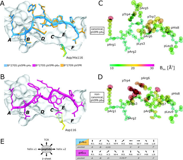
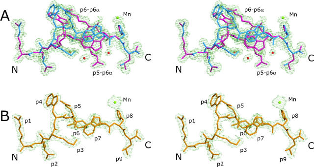
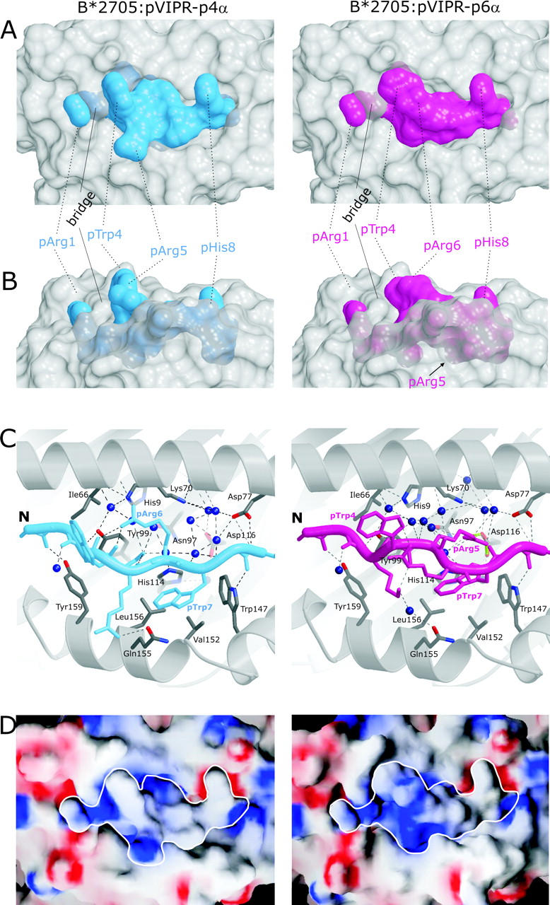
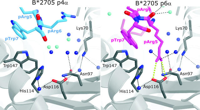
The products of the human leukocyte antigen subtypes HLA-B*2705 and HLA-B*2709 differ only in residue 116 (Asp vs. His) within the peptide binding groove but are differentially associated with the autoimmune disease ankylosing spondylitis (AS); HLA-B*2705 occurs in AS-patients, whereas HLA-B*2709 does not. The subtypes also generate differential T cell repertoires as exemplified by distinct T cell responses against the self-peptide pVIPR (RRKWRRWHL). The crystal structures described here show that pVIPR binds in an unprecedented dual conformation only to HLA-B*2705 molecules. In one binding mode, peptide pArg5 forms a salt bridge to Asp116, connected with drastically different interactions between peptide and heavy chain, contrasting with the second, conventional conformation, which is exclusively found in the case of B*2709. These subtype-dependent differences in pVIPR binding link the emergence of dissimilar T cell repertoires in individuals with HLA-B*2705 or HLA-B*2709 to the buried Asp116/His116 polymorphism and provide novel insights into peptide presentation by major histocompatibility antigens.


Figures

Figure 1.
Figure 1.
pVIPR conformations, atomic displacement ellipsoids, and B factors. (A) Superimposition of the canonical pVIPR conformations (p4α) found in B*2705 (blue) and B*2709 (gold). (B) The noncanonical pVIPR conformation (p6α, pink) observed only in B*2705. The peptides are viewed from the side of the α2 helix together with a molecular surface covering the floor and back of the binding groove. The subtype-specific residue 116 is indicated also (Asp116, yellow; His116, turquoise); the bidentate salt bridge to Asp116 is drawn with green dotted lines in B. The binding pockets A–F are shown in bold letters. Atomic displacement ellipsoids for pVIPR-p4α and -p6α in C and D are colored according to the equivalent isotropic temperature factors B (Å2) (see color bar). (E, left) Schematic description of side chain orientation when looking from the NH2 to the COOH terminus of pVIPR. Bottom of peptide binding groove indicated by “β-sheet” and side for T cell recognition by “TCR”. (E, right) The orientation of the peptide side chains in the p4α and p6α conformations as in E (left). The respective binding pockets (A–F) are indicated as well. It is clear from this representation and Fig. 1 (A and B) that the two pVIPR conformations show major differences only from pLys3 to pTrp7.
Figure 2.
Figure 2.
Final electron density of pVIPR conformations in B*2705 and B*2709. Stereo images of the final 2Fo-Fc electron density contoured at 1 σ level, displayed in light green. The two B*2705:pVIPR conformations are shown in A, the respective B*2709 complex is shown in B. The peptides are color coded as in Fig. 1 (A and B): water molecules as red and Mn2+ as green spheres.
Figure 3.
Figure 3.
Molecular surfaces and contacts of pVIPR in the p4α and p6α conformations. (A and B) Molecular surfaces show the central part of the B*2705 peptide binding groove in gray and the pVIPR peptide in the p4α and p6α conformations (color coded as in Fig. 1 [A and B]). The binding groove has been rendered semi-transparent, allowing also the inspection of buried side chains exhibiting conformational differences. In A, the view is TCR-like, straight onto the peptide, whereas in B (rotated by 90° about a horizontal axis), the view is through the α2 helix. The center section of the peptide shows clear shape differences between the p4α and p6α conformations. (C) pVIPR hydrogen bonding in p4α and p6α, color coded as in Fig 1 (A and B). Only side chains with different binding modes (residues p3–p7) are shown. The binding groove's secondary structure is represented as gray spirals (α helices) and arrows (β strands) together with selected interacting residues (carbon atoms, gray; oxygens, red; and nitrogens, blue). Hydrogen bonds are depicted as black broken lines, the pArg5–Asp116 bidentate salt bridge is depicted as green dotted lines, and water molecules are depicted as dark blue spheres. (D) Electrostatic surfaces of both pVIPR conformations. Red indicates negative, blue indicates positive surface charge, and gray areas are uncharged. The view is looking straight onto the binding groove as in A. The border of the peptides is highlighted in white.
Figure 4.
Figure 4.
Conformation-dependent peptide contacts with F-pocket residues. The F-pocket architecture and intermolecular interactions in B*2705:pVIPR-p4α (left) and -p6α (right), with relevant part of the peptide shown (same color code as in Fig. 1 [A and B]). Fully occupied water molecules are shown in dark blue and partially occupied ones (related to a specific peptide conformation) are in turquoise. The space occupied by pArg5-p6α is filled by water molecules in the p4α binding mode. The view is looking along the binding groove with the peptide COOH terminus in front.

References

    1. Madden, D.R. 1995. The three-dimensional structure of peptide-MHC complexes. Annu. Rev. Immunol. 13:587–622. - PubMed
    1. Saper, M.A., P.J. Bjorkman, and D.C. Wiley. 1991. Refined structure of the human histocompatibility antigen HLA-A2 at 2.6 A resolution. J. Mol. Biol. 219:277–319. - PubMed
    1. Matsumura, M., D.H. Fremont, P.A. Peterson, and I.A. Wilson. 1992. Emerging principles for the recognition of peptide antigens by MHC class I molecules. Science. 257:927–934. - PubMed
    1. Benjamin, R., and P. Parham. 1990. Guilt by association: HLA-B27 and ankylosing spondylitis. Immunol. Today. 11:137–142. - PubMed
    1. Szpak, Y., J.C. Vieville, T. Tabary, M.C. Naud, M. Chopin, C. Edelson, J.H. Cohen, J. Dausset, Y. de Kozak, and M. Pla. 2001. Spontaneous retinopathy in HLA-A29 transgenic mice. Proc. Natl. Acad. Sci. USA. 98:2572–2576. - PMC - PubMed

Publication types