. 2004 Jan 19;199(2):283-93.
doi: 10.1084/jem.20030648.

HIV-1 cell to cell transfer across an Env-induced, actin-dependent synapse

Affiliations

HIV-1 cell to cell transfer across an Env-induced, actin-dependent synapse

Clare Jolly et al. J Exp Med. .

Abstract

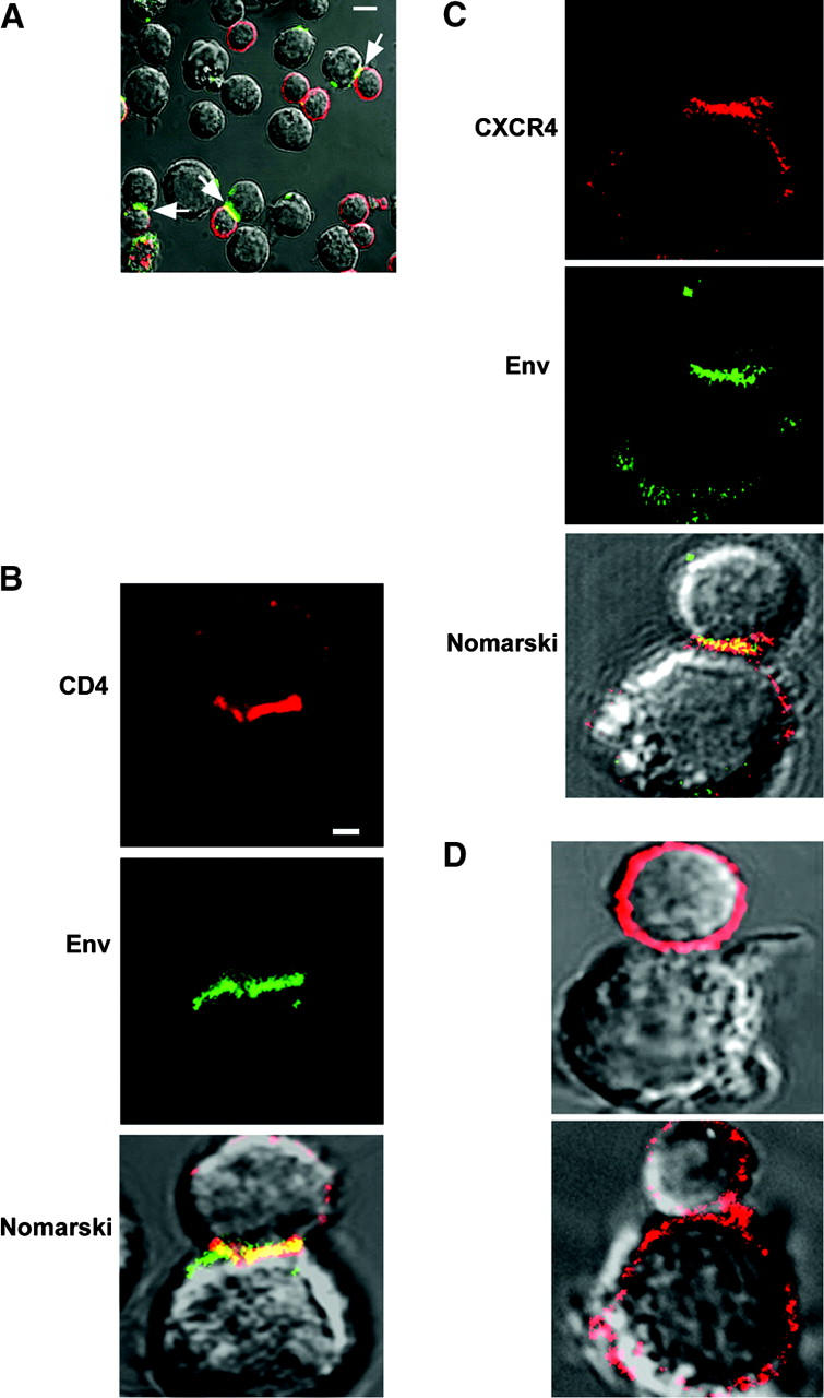
Direct cell-cell transfer is an efficient mechanism of viral dissemination within an infected host, and human immunodeficiency virus 1 (HIV-1) can exploit this mode of spread. Receptor recognition by HIV-1 occurs via interactions between the viral surface envelope glycoprotein (Env), gp120, and CD4 and a chemokine receptor, CCR5 or CXCR4. Here, we demonstrate that the binding of CXCR4-using HIV-1-infected effector T cells to primary CD4(+)/CXCR4(+) target T cells results in rapid recruitment to the interface of CD4, CXCR4, talin, and lymphocyte function-associated antigen 1 on the target cell, and of Env and Gag on the effector cell. Recruitment of these membrane molecules into polarized clusters was dependent on Env engagement of CD4 and CXCR4 and required remodelling of the actin cytoskeleton. Transfer of Gag from effector to target cell was observed by 1 h after conjugate formation, was independent of cell-cell fusion, and was probably mediated by directed virion fusion with the target cell. We propose that receptor engagement by Env directs the rapid, actin-dependent recruitment of HIV receptors and adhesion molecules to the interface, resulting in a stable adhesive junction across which HIV infects the target cell.


Figures

Figure 1.
Figure 1.
Polarization of HIV-1 receptors and Env to the interface. Conjugates formed for 30 min between JurkatLAI (effector) and primary CD4+ T (target) cells were labeled by indirect immunofluorescence, fixed, and analyzed by confocal microscopy. Images are single two-dimensional x-y sections through the central region of the cells with the corresponding Nomarski view of the cell surface. (A) Cells were stained during conjugate formation for CD4 (red) and gp41 (green) with mAbs L120 and 50-69, respectively. Conjugates with receptor-Env polarization and colocalization (yellow) are indicated with arrows. Bar, 10 μm. (B) Higher magnification of a target–effector cell conjugate showing polarized CD4 (red) and Env (green) colocalized (yellow) at the interface. Bar, 1 μm. (C) CXCR4 (red) on the target and effector cells copolarizes with Env (green) on the effector cell. (D) Conjugates between target cells and uninfected Jurkats do not show polarization of CD4 (top) or CXCR4 (bottom).
Figure 2.
Figure 2.
Talin and LFA-1 cluster at the target–effector cell interface. Conjugates between effector and target cells formed for 1 h at 37°C were stained for Env with 50-69 (green). The target cell was identified using a CD4-specific rabbit polyclonal serum (staining not depicted) and is indicated with an arrow. (A) Conjugates were either fixed with prechilled methanol and stained for the actin adaptor protein talin (red), (B) stained for the integrin LFA-1, or (C) CD3 before fixing in formaldehyde.
Figure 3.
Figure 3.
Kinetics of receptor polarization. Target and effector cells were mixed and incubated for 30 min on ice to synchronize cell adhesion to the poly-l-lysine–coated coverslips. Coverslips were transferred to 37°C and incubated for 10, 30, or 60 min before transfer to ice with the addition of NaN3-containing buffer. Cells were either stained during conjugate formation with mAbs to Env and CD4 or to Env alone followed by incubation on ice to detect CD4 (using rabbit polyclonal serum), CXCR4, and LFA-1. Conjugates were fixed, stained with secondary antibodies, and analyzed by confocal microscopy. Conjugates were scored for copolarization of Env and CD4 (black bars), Env and LFA-1 (white bars), or polarization of CXCR4 alone (diagonally striped bars) The error bars show the standard error of the mean.
Figure 4.
Figure 4.
Receptor clustering requires the actin cytoskeleton. (A) Effector target cell conjugates were incubated at 37°C for 1 h in the presence of mAbs for CD4 (red) and Env (green). Conjugates were fixed, permeabilized, and stained for actin (blue). Clusters of f-actin on the target cell are indicated with arrows. (B) Target cells were treated with 1 μM of cytochalasin D (top) or jasplakinolide (bottom) for 1 h at 37°C. Treated cells were washed and incubated with effector cells at 37°C for 1 h with mAbs against CD4 (red) and Env (green).
Figure 4.
Figure 4.
Receptor clustering requires the actin cytoskeleton. (A) Effector target cell conjugates were incubated at 37°C for 1 h in the presence of mAbs for CD4 (red) and Env (green). Conjugates were fixed, permeabilized, and stained for actin (blue). Clusters of f-actin on the target cell are indicated with arrows. (B) Target cells were treated with 1 μM of cytochalasin D (top) or jasplakinolide (bottom) for 1 h at 37°C. Treated cells were washed and incubated with effector cells at 37°C for 1 h with mAbs against CD4 (red) and Env (green).
Figure 5.
Figure 5.
Polarization and transfer of HIV antigens. Conjugates were formed between effector and target cells in the presence of mAbs specific for CD4 (green) and Env (blue). (A) Cells were fixed, permeabilized, and stained for Gag (red); arrows show Gag staining within the target cell. (B) Conjugates were prepared as for A, but incubated at 37°C for 3 h before fixation, permeabilization, and Gag staining. (C) Conjugates were prepared for 1 h as aforementioned, except that the effector cells were prelabeled with CellTracker green. After permeabilization, cells were stained for Gag (blue) and EEA1 (red); a three-dimensional reconstruction of a z series of images is shown.
Figure 5.
Figure 5.
Polarization and transfer of HIV antigens. Conjugates were formed between effector and target cells in the presence of mAbs specific for CD4 (green) and Env (blue). (A) Cells were fixed, permeabilized, and stained for Gag (red); arrows show Gag staining within the target cell. (B) Conjugates were prepared as for A, but incubated at 37°C for 3 h before fixation, permeabilization, and Gag staining. (C) Conjugates were prepared for 1 h as aforementioned, except that the effector cells were prelabeled with CellTracker green. After permeabilization, cells were stained for Gag (blue) and EEA1 (red); a three-dimensional reconstruction of a z series of images is shown.
Figure 6.
Figure 6.
Kinetics of Env-mediated cell–cell fusion. Effector (red) and target (green) cells were labeled with cytoplasmic dyes, and conjugates were formed at 37°C in the presence or absence of DP178 for various times. The cells were fixed, and the conjugates were analyzed for fusion and mixing of cytoplasmic dyes (yellow). After 3 h of conjugate formation (left), no transfer of dye is detected; by 24 h, transfer of dye is apparent (middle) and this is blocked in conjugates formed for 24 h in the presence of DP178 (right).
Figure 7.
Figure 7.
Immunoelectron microscopy showing clusters of HIV-1 particles at the synapse. Effector–target conjugates were formed for 1 h and processed for electron microscopy. (A) Low magnification (11,000×) of an ultrathin section through an effector–target cell conjugate. Closely apposed regions of membrane are numbered (1 and 2). Bar, 1 μm. (B) Higher magnification (30,000×) of the interface in the same conjugate. Small arrows highlight 6 nm of gold particles bound to CD4 on the target cell. Large arrows show mature virions associated with the membrane of the target cell, and the asterisk marks a structure resembling a budding virion. Bar, 300 nm.

References

    1. Johnson, D., and M. Huber. 2002. Directed egress of animal viruses promotes cell-to-cell spread. J. Virol. 76:1–8. - PMC - PubMed
    1. Igakura, T., J. Stinchcombe, P. Goon, G. Taylor, J. Weber, G. Griffiths, Y. Tanaka, M. Osame, and C. Bangham. 2003. Spread of HTLV-1 between lymphocytes by virus-induced polarisation of the cytoskeleton. Science. 299:1713–1716. - PubMed
    1. Phillips, D. 1994. The role of cell-to-cell transmission in HIV infection. AIDS. 8:719–731. - PubMed
    1. Haase, A. 1999. Population biology of HIV-1 infection: viral and CD4+ T cell demographics and dynamics in lymphatic tissue. Annu. Rev. Immunol. 17:625–656. - PubMed
    1. Sato, H., J. Orenstein, D. Dimitrov, and M. Martin. 1992. Cell-cell spread of HIV-1 occurs within minutes and may not involve the participation of virus particles. Virology. 186:712–724. - PubMed

Publication types

MeSH terms