. 2004 Jan 19;164(2):243-53.
doi: 10.1083/jcb.200309096.

A divergent canonical WNT-signaling pathway regulates microtubule dynamics: dishevelled signals locally to stabilize microtubules

Affiliations

A divergent canonical WNT-signaling pathway regulates microtubule dynamics: dishevelled signals locally to stabilize microtubules

Lorenza Ciani et al. J Cell Biol. .

Abstract

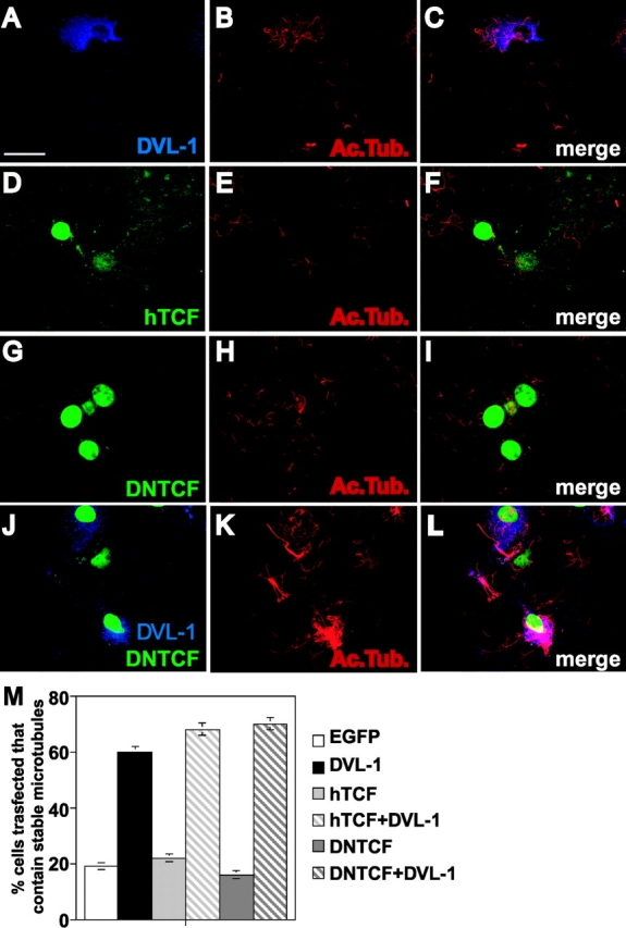
Dishevelled (DVL) is associated with axonal microtubules and regulates microtubule stability through the inhibition of the serine/threonine kinase, glycogen synthase kinase 3beta (GSK-3beta). In the canonical WNT pathway, the negative regulator Axin forms a complex with beta-catenin and GSK-3beta, resulting in beta-catenin degradation. Inhibition of GSK-3beta by DVL increases beta-catenin stability and TCF transcriptional activation. Here, we show that Axin associates with microtubules and unexpectedly stabilizes microtubules through DVL. In turn, DVL stabilizes microtubules by inhibiting GSK-3beta through a transcription- and beta-catenin-independent pathway. More importantly, axonal microtubules are stabilized after DVL localizes to axons. Increased microtubule stability is correlated with a decrease in GSK-3beta-mediated phosphorylation of MAP-1B. We propose a model in which Axin, through DVL, stabilizes microtubules by inhibiting a pool of GSK-3beta, resulting in local changes in the phosphorylation of cellular targets. Our data indicate a bifurcation in the so-called canonical WNT-signaling pathway to regulate microtubule stability.


Figures

Figure 1.
Figure 1.
DVL-1 mimics the axonal remodeling activity of WNTs. (A) DVL-1 expression (asterisk) induces growth cone enlargement and increased axonal diameter, and decreases axon length (filled arrowheads) compared with untransfected differentiated neurons from NB2a cells (empty arrowhead). (B) Control neurons have thin axons with relatively small growth cones and few stable microtubules. (C) Expression of DVL-1 increases the number of stable microtubules in axons and induces looping and unbundling of stable micro- tubules at enlarged growth cones. (D and E) Expression of DVL-1 induces the translocation of endogenous β-catenin into the nucleus (arrowheads). Dashed lines show the shape of the growth cone. Bars: (A, D, and E) 15 μm, (B and C) 2 μm.
Figure 2.
Figure 2.
TCF is not required for DVL microtubule-stabilizing function. (A–C) Expression of DVL-1 protects microtubule against nocodazole. (D–I) Neither hTCF nor ΔNTCF protect microtubules against nocodazole. (J and L) Expression of ΔNTCF in DVL-expressing cells does not affect the ability of DVL to stabilize microtubules against nocodazole. Bar, 50 μm. (M) Graph shows the percentage of cells containing microtubules after nocodazole treatment. Values are mean ± SEM from three different experiments (at least 30 neurons were counted in each experiment).
Figure 3.
Figure 3.
β-catenin is dispensable for DVL-stabilizing function. (A–C) Expression of wild-type β-catenin does not stabilize microtubules against nocodazole. (D–G) Mutant β-catenin (ARM β-cat) lacking the transactivation domain does not interfere with the ability of DVL to stabilize microtubules. Bar, 50 μm. (H) Graph shows the percentage of cells containing microtubules after nocodazole treatment. In the first three columns, the error bars are too small to be seen. Values are mean ± SEM from three different experiments (at least 70 neurons were counted in each experiment).
Figure 4.
Figure 4.
The microtubule-stabilizing function of DVL does not require transcriptional activity. (A) Schematic representation of the experimental design. (B–D) Cells expressing DVL-ER in the absence of β-estradiol do not have stable microtubules when treated with nocodazole and actinomycin D. (E–G) After β-estradiol, DVL-ER–expressing cells contain nocodazole-resistant microtubules, even in the presence of actinomycin D. Bar, 50 μm.
Figure 5.
Figure 5.
Local effect of DVL microtubule-stabilizing function. (A) In the absence of nocodazole and β-estradiol, DVL-ER is evenly distributed in NB2a differentiated neurons. (B) A significant amount of DVL-ER has a punctate distribution in the cell body after 1 h β-estradiol induction. (C) DVL-ER is present at significant levels along the axon after 5 h induction with β-estradiol. (D and D′) In the presence of nocodazole but in the absence of β-estradiol, neurons lose their microtubules. (E and E′) After 1 h of β-estradiol, microtubules are stabilized mainly in the cell body after nocodazole. (F and F′) After 5 h of β-estradiol induction, a significant number of neurons contain axonal microtubules after nocodazole treatment, coinciding with the presence of DVL along the axon (arrowheads) Bar, 15 μm. (G) Quantification of the percentage of DVL-expressing cells that have stable microtubules in the cell body and axon under different periods of β-estradiol treatment. (H) Graph shows the correlation between the localization of DVL-ER in axons and the presence of stable microtubule along the axons. Values are mean ± SEM from three experiments (at least 70 neurons were counted in each experiment).
Figure 6.
Figure 6.
DVL stabilizes microtubule via inhibition of GSK-3β without the requirement of FRAT. (A and B) NB2a neurons treated with lithium show a significant increase in the number of stable microtubules after nocodazole treatment when compared with control NaCl-treated neurons. (C) Graph shows the percentage of neurons with stable microtubules after nocodazole treatment. Note the proportional increase in microtubule stability with increased concentrations of lithium. (D–F) Full-length FRAT does not protect microtubules against nocodazole. (G–J) Expression of ΔCFRAT, lacking the GSK-3β binding domain, does not affect the ability of DVL-1 to protect microtubules against nocodazole (arrows). Bars, 15 μm.
Figure 7.
Figure 7.
Axin associates with microtubules and stabilizes microtubules through DVL. (A) Endogenous Axin comes down with brain microtubule pellets obtained through one and two rounds of polymerization and repolymerization. (B) Endogenous Axin is associated with microtubules along the axon (filled arrowhead) and at growth cones (empty arrowhead) of cerebellar granule cell neurons. (C) Expression of DVL in COS7 cells does not change the level of endogenous Axin or GSK-3β associated with microtubules. (D–F) Expression of DVL or Axin in NB2a neurons protects microtubules from nocodazole. (G) Neurons expressing both Axin and ΔPDZ-DVL lose their microtubules after nocodazole treatment (open arrowheads), whereas cells expressing Axin alone (filled arrowheads) retain their microtubules. (H) Fewer cells expressing AxinL-P carrying a mutation in the GSK-3β binding site exhibit microtubule stability (arrowheads). Bar, 10 μm. (I) Graph shows that DVL and Axin have similar levels of microtubule stabilizing function. Expression of both DVL and Axin do not have an additive effect. AxL-P and ΔDIX-Axin exhibit a lower microtubule-stabilizing function when compared with wild-type Axin. Full-length DVL rescues both mutant forms of Axin. In contrast, ΔPDZ-DVL has a low level of microtubule stabilizing function when compared with full length DVL. Expression of Axin was unable to rescue microtubule stability in cells expressing ΔPDZ-DVL. Values are mean ± SEM.
Figure 8.
Figure 8.
DVL stabilizes microtubules in primary neurons and down-regulates the level of GSK-3β–mediated phosphorylation of MAP-1B in developing axons. (A–F) Cerebellar granule cell neurons expressing DVL-1 contain stable microtubules (filled arrowheads), whereas EGFP-expressing control neurons lose their microtubules after nocodazole treatment (open arrowheads). (G–I) Cerebellar granule cell neurons expressing EGFP exhibit a significant level of GSK-3β–phosphorylated MAP-1B-P along the axon with similar levels to untransfected neurons (open arrowheads). (J–L) Expression of DVL-1 in cerebellar granule cell neurons decreases the level of GSK-3β–phosphorylated MAP-1B-P along the axon (filled arrowheads). Bar, 10 μm.

References

    1. Andersen, S.S., and G.Q. Bi. 2000. Axon formation: a molecular model for the generation of neuronal polarity. Bioessays. 22:172–179. - PubMed
    1. Arias, A.M., A.M. Brown, and K. Brennan. 1999. Wnt signalling: pathway or network? Curr. Opin. Genet. Dev. 9:447–454. - PubMed
    1. Baas, P.W. 1999. Microtubules and neuronal polarity: lessons from mitosis. Neuron. 22:23–31. - PubMed
    1. Behrens, J., B.A. Jerchow, M. Wurtele, J. Grimm, C. Asbrand, R. Wirtz, M. Kuhl, D. Wedlich, and W. Birchmeier. 1998. Functional interaction of an axin homolog, conductin, with β-catenin, APC, and GSK3β. Science. 280:596–599. - PubMed
    1. Bosc, C., J.D. Cronk, F. Pirollet, D.M. Watterson, J. Haiech, D. Job, and R.L. Margolis. 1996. Cloning, expression, and properties of the microtubule-stabilizing protein STOP. Proc. Natl. Acad. Sci. USA. 93:2125–2130. - PMC - PubMed

Publication types

MeSH terms