. 2004 Feb 3;101(5):1189-94.
doi: 10.1073/pnas.0307966100. Epub 2004 Jan 20.

A proton-coupled dynamic conformational switch in the HIV-1 dimerization initiation site kissing complex

Affiliations

A proton-coupled dynamic conformational switch in the HIV-1 dimerization initiation site kissing complex

Mihaela-Rita Mihailescu et al. Proc Natl Acad Sci U S A. .

Abstract

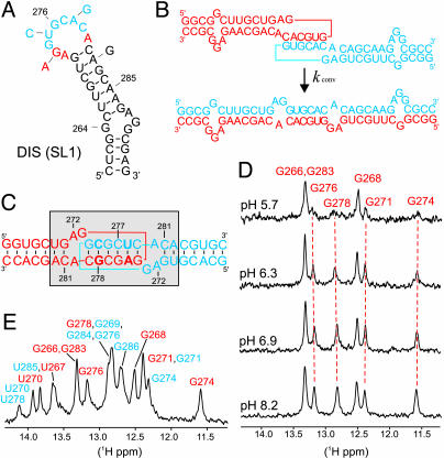
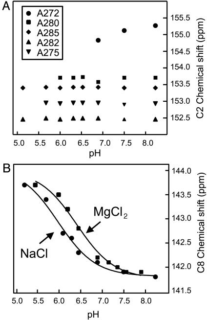
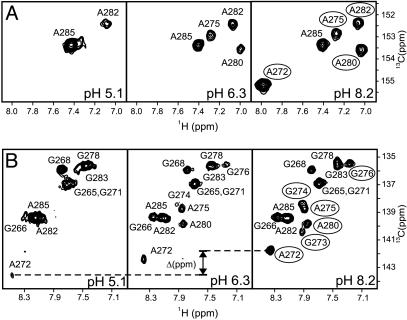
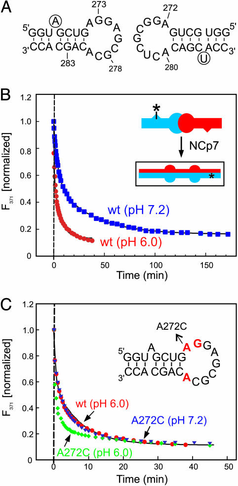
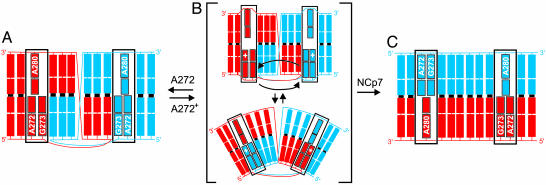
In HIV type 1 (HIV-1), the dimerization initiation site (DIS) is the sequence primarily responsible for initiating the noncovalent linkage of two homologous strands of genomic RNA during viral assembly. The DIS loop contains an autocomplementary hexanucleotide sequence and forms a symmetric homodimer through a loop-loop kissing interaction. In a structural rearrangement catalyzed by the HIV-1 nucleocapsid protein (NCp7) and suggested to be associated with maturation of the budded viral particle, the DIS converts from a metastable kissing dimer to an extended duplex. Here, we demonstrate that the DIS kissing dimer displays localized conformational dynamics that result from the specific protonation of the N1 base nitrogen of the DIS loop residue A272 at near-physiological pH. The rate of NCp7-catalyzed maturation of the DIS kissing dimer is also shown to directly correlate with the observed proton-coupled conformational dynamics, where NCp7 is found to convert the dynamic A272 protonated state with a faster rate. Taken together, these results reveal a previously undescribed role for base protonation in modulating local RNA structure and demonstrate a mechanism for promoting the chaperone-mediated structural rearrangement of a kinetically trapped RNA conformational state.


Figures

Fig. 1.
Fig. 1.
Secondary structure and pH-dependent DIS dynamics. (A) Stem–loop sequence and secondary structure for DIS (SL1) from HIV-1 subtype A. The autocomplementary hexanucleotide loop sequence is shown in blue, and conserved purines are bolded in red. Arrows indicate differences in sequence from the HIV-1 subtype B strain. (B) Representation of the DIS homodimer conversion from kissing dimer to extended duplex, with DIS stem–loops shown in blue and red; purine junction bases are boxed. (C) Sequence and secondary structure of the DIS23(GA)·DIS23(HxUC) heterodimeric kissing complex formed by DIS23(GA) in red and DIS23(HxUC) designed with stem sequences noncomplementary to the stems of DIS23(GA) in blue. Point mutation palindromic hexanucleotide DIS loop sequences used to generate heterodimeric complexes are bolded. The conformationally dynamic region is shown by a shaded box. (D)1D 15N-filtered 1H-NMR spectra of the imino proton region of a 13C,15N-A,G-labeled DIS23(GA)·DIS23(HxUC) kissing complex in NMR buffer and 300 mM NaCl as a function of pH. Imino assignments are indicated and the subset of pH-dependent resonances followed by dashed red lines. (E) Assigned imino proton resonances [DIS23(GA) in red and DIS23(HxUC) in blue] of the DIS kissing dimer at pH 8.2.
Fig. 2.
Fig. 2.
In situ determination of a pKa for A272. (A) Plot of the C2 carbon chemical shifts vs. pH of the five adenosines in the DIS23(GA) stem–loop in the 13C,15N-A,G-labeled DIS23(GA)·DIS23(HxUC) kissing complex formed in the presence of 300 mM NaCl. Note that resonances from residue A272, A280, and A275 become exchange broadened beyond detection as the pH is lowered, and, therefore, the pH-dependent correlation of the C2 chemical shifts for these bases is incomplete. (B) Plot of the C8 carbon chemical shift of A272 from DIS23(GA) vs. pH in the 13C,15N-A,G-labeled DIS23(GA)·DIS23(HxUC) kissing complex formed in the presence of either 300 mM NaCl or 3.3 mM MgCl2. Solid lines are best fits using a Henderson–Hasselbach equation.
Fig. 3.
Fig. 3.
Selected regions of a 600-MHz 2D 1H-13C HSQC spectrum of the 13C,15N-A,G-labeled DIS23(GA)·DIS23(HxUC) kissing complex formed in NMR buffer supplemented with 300 mM NaCl showing purine C2-H2 (A) and C8-H8 (B) correlations at pH 5.1, 6.3, and 8.2. Note that, although the C2 chemical shift is expected to shift upfield by as much as 8 ppm on protonation of an adenosine base, no C2–H2 crosspeaks are observed outside the plotted region over the entire pH range. The change in C8 carbon chemical shift for A272 is indicated; other resolved crosspeaks observed to broadened at low pH are circled in the pH 8.2 spectrum.
Fig. 4.
Fig. 4.
pH dependence of NCp7-catalyzed DIS maturation. (A) Sequence of the DIS24(GA)-4ap and DIS24(UC) stem–loops used in the fluorescence experiments. 2-AP label at position 4 of DIS24(GA)-4ap and the added U-bulge in DIS24(UC) are circled. (B) Plot of the normalized fluorescence decay as a function of time after NCp7 protein was added to a DIS24(GA)-4ap·DIS24(UC) kissing complex preformed in NMR buffer with 5 mM MgCl2 at pH 6.0 (red circles) and 7.2 (blue squares). Structural isomerization rates (pH 6.0: kconv = 1.11 ± 0.12 min–1, karr = 0.11 ± 0.01 min–1; pH 7.2: kconv = 0.23 ± 0.01 min–1, karr = 0.026 ± 0.001 min–1) were fit by using a double exponential rate equation. (Inset) A schematic of the NCp7-catalyzed structural isomerization of the kissing dimer. Asterisks indicate the position of 2-AP in the DIS24(GA)-4ap stem-loop. (C) Plot of the normalized fluorescence decay as a function of time after NCp7 protein was added to kissing complexes formed with A272C mutant DIS sequences DIS24(GA,A272C)-4ap·DIS24(UC,A272C) by using conditions as in A, at pH 6.0 (green diamonds) and 7.2 (blue triangles). Structural isomerization rates (pH 6.0: kconv = 4.92 ± 0.59 min–1, karr = 0.28 ± 0.03 min–1; pH 7.2: kconv = 3.94 ± 0.49 min–1, karr = 0.22 ± 0.01 min–1) were fit by using a double exponential rate equation. The DIS wild-type sequence data (red circles) is shown for comparison. (Inset) A schematic of the DIS24(GA)-4ap stem–loop with junction bases shown in red and the A272C mutation indicated.
Fig. 5.
Fig. 5.
Model for the pH-dependent structural interconversion of the DIS dimer. DIS stem–loops are shown in blue and red to the second base in the stem. Junction bases are labeled and boxed. (A) The A272 nonprotonated state of the DIS kissing dimer observed at high pH. (B) A two-state representation of the dynamic ensemble of states that results on protonation of A272. Black arrows indicate possible 5′ base motions of A272 and G273 that could result in the observed exchange broadening of the NMR resonances belonging to these and flanking residues. Note that the ensemble may also be comprised of states in which only one of the 5′ bases flips between stacked positions or states in which the purine base motions are more localized. (C) The mature duplex dimer state, showing a model of the NMR-determined structure (39, 40) with all junction bases stacked in toward the helix.

Similar articles

Cited by

References

    1. Skripkin, E., Paillart, J. C., Marquet, R., Ehresmann, B. & Ehresmann, C. (1994) Proc. Natl. Acad. Sci. USA 91, 4945–4949. - PMC - PubMed
    1. Laughrea, M. & Jette, L. (1994) Biochemistry 33, 13464–13474. - PubMed
    1. Paillart, J. C., Marquet, R., Skripkin, E., Ehresmann, C. & Ehresmann, B. (1996) Biochimie 78, 639–653. - PubMed
    1. Berkhout, B. & vanWamel, J. L. B. (1996) J. Virol. 70, 6723–6732. - PMC - PubMed
    1. Clever, J. L. & Parslow, T. G. (1997) J. Virol. 71, 3407–3414. - PMC - PubMed

Publication types

LinkOut - more resources