. 2004 Feb 3;101(5):1241-6.
doi: 10.1073/pnas.0307708100. Epub 2004 Jan 20.

Histone deacetylase (HDAC) inhibitor activation of p21WAF1 involves changes in promoter-associated proteins, including HDAC1

Affiliations

Histone deacetylase (HDAC) inhibitor activation of p21WAF1 involves changes in promoter-associated proteins, including HDAC1

C-Y Gui et al. Proc Natl Acad Sci U S A. .

Abstract

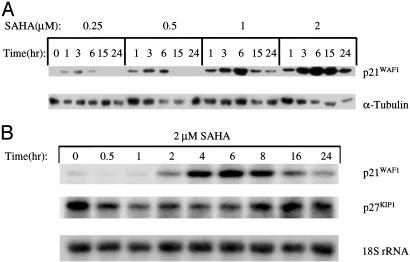
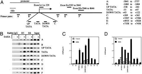
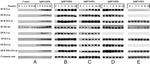
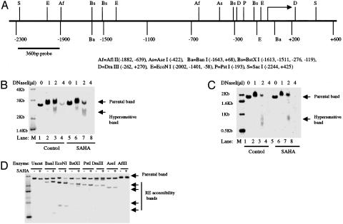
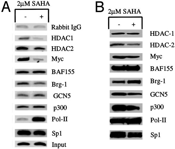
Histone deacetylase (HDAC) inhibitors (HDACi) cause cancer cell growth arrest and/or apoptosis in vivo and in vitro. The HDACi suberoylanilide hydroxamic acid (SAHA) is in phase I/II clinical trials showing significant anticancer activity. Despite wide distribution of HDACs in chromatin, SAHA alters the expression of few genes in transformed cells. p21(WAF1) is one of the most commonly induced. SAHA does not alter the expression of p27(KIPI), an actively transcribed gene, or globin, a silent gene, in ARP-1 cells. Here we studied SAHA-induced changes in the p21(WAF1) promoter of ARP-1 cells to better understand the mechanism of HDACi gene activation. Within 1 h, SAHA caused modifications in acetylation and methylation of core histones and increased DNase I sensitivity and restriction enzyme accessibility in the p21(WAF1) promoter. These changes did not occur in the p27(KIPI) or epsilon-globin gene-related histones. The HDACi caused a marked decrease in HDAC1 and Myc and an increase in RNA polymerase II in proteins bound to the p21(WAF1) promoter. Thus, this study identifies effects of SAHA on p21(WAF1)-associated proteins that explain, at least in part, the selective effect of HDACi in altering gene expression.


Figures

Fig. 1.
Fig. 1.
SAHA rapidly increases the p21WAF1 mRNA and protein levels in ARP-1 cells. (A) ARP-1 cells were cultured with the concentrations of SAHA indicated and for the times indicated. Whole-cell extracts from 107 ARP-1 cells were prepared, and 30 μg of the extract protein was loaded in SDS/15% PAGE gel and immunoblotted with antibody against p21WAF1 protein. A duplicate gel and membrane were blotted with the antibody for α-tubulin. (B) Total cellular RNA was prepared from ARP-1 cells cultured with 2 μM SAHA for the times indicated; 10 μg of RNA was electrophoresed on a 1% formaldehyde-denatured agarose gel and then transferred to a Hybond N+ nylon membrane. The membrane was hybridized with a 32P-labeled 695-bp p21WAF1 cDNA probe, then stripped and rehybridized with a 32P-labeled 383-bp PstI-digested p27KIP1 cDNA probe and a 32P-labeled 50-mer 18S rRNA oligonucleotide probe sequentially.
Fig. 2.
Fig. 2.
Effect of SAHA on acetylation of p21WAF1-associated histone H3 and H4. (A) A schematic graph of the p21WAF1 gene indicating the location of nine pairs of primers used for PCR amplification in ChIP assays. The position of the primer pairs designated A, B, UP-TATA, TATA, DOWN-TATA, C, D, E, and F corresponded to the base pairs indicated from upstream (–3,841 bp) to downstream (+7,438 bp) of the transcription initiation site. (B) ChIP assays were prepared by using rabbit normal IgG, anti-diacetylated H3 (K9/14-ac), and anti-tetraacetylated H4 (K5/8/12/16-ac). The samples were amplified with each of the nine pairs of primers. ARP-1 cell were cultured without (–) and with (+) 2 μM SAHA for 3 h. (C) p21WAF1 DNA corresponding to the indicated probe as a percent of the input DNA associated with histone H3. (D) p21WAF1 DNA corresponding to indicated probe as a percent of the input DNA associated with acetylated histone H4.
Fig. 3.
Fig. 3.
SAHA-induced modifications of specific lysine residues of histone H3 and H4. (A) ARP-1 cells (2.5 × 10⁵ cells per ml) were cultured without or with 2 μM SAHA for the indicated times. Histones were prepared and analyzed as detailed in Experimental Procedures. The primer histone lysine and serine antibodies used for Western blot analysis are indicated. (B) ChIP analyses of histone H3 and H4 modifications in p21WAF1 proximal promoter region (TATA box primer pair, –52 to +28) of ARP-1 cells at zero time and cultured with 2 μM SAHA for the times indicated. One set of ChIP analyses was performed with the antibodies indicated. (C) ChIP analyses of histone H3 and H4 modifications in p21WAF1 distal promoter region (primer pair B, –1,703 to –1,623) of ARP-1 cells used for B.(D) ChIP analyses of histone modifications in p27KIP1 promoter (from –1,320 to –1,412) prepared from ARP-1 cells used for B and C. The antibodies were used for ChIP assays as for AC except for anti-H3 K9 mc, H4 K5 ac, and H4-K 16 ac. (E) ChIP analyses of histones modification in ε-globin promoter (see Experimental Procedures) prepared from ARP-1 cells used in BD. No signal was detected with antibody to H3, K9ac.
Fig. 4.
Fig. 4.
DNase I sensitivity and restriction enzyme accessibility of the p21WAF1 gene from ARP-1 cells cultured with SAHA. (A) A schematic graph of p21WAF1 promoter region indicating the recognition sites of the restriction enzymes used for accessibility assay and the location of the probe used for Southern blot hybridization (360 bp of SacI-to-AflII fragment). Restriction enzymes are represented with a single letter or two letters, and the recognition sites are shown as numbers with reference to the transcriptional start site as +1. The subscript identifies the restriction enzymes. (B) DNase I sensitivity assay of the p21WAF1 promoter of ARP-1 cells cultured with or without 2 μM SAHA for 3 h. “Parental band” is the SacI fragment of p21WAF1 promoter (2,669 bp; –2,244 bp to 4,425 bp). “DNase hypersensitive” band is a smear (2.2–2.4 kb). Lane M, 32P-labeled 1-kb DNA ladder (Invitrogen). Lanes 1–4, nuclei of ARP-1 cells digested with increasing DNase I (Invitrogen, 105 units/μl) volume (0, 1, 2, and 4 μl of DNase I, respectively) for 10 min at room temperature. Lanes 5–8, nuclei of ARP-1 cells cultured with 2 μM SAHA for 3 h; the nuclei were digested with increasing DNase I volume (0, 1, 2, and 4 μl of DNase I, respectively) for 10 min at room temperature. (C) DNase I hypersensitivity assay of p27KIP1 promoter of ARP-1 cells with or without the treatment of 2 μM SAHA for 3 h. The analyses were preformed as indicated for B. (D) Restriction enzyme accessibility assays of chromatin of p21WAF1 promoter of ARP-1 cells cultured without (–) or with 2 μM SAHA for 3 h. Nuclei were prepared and digested with AflII, AseI, BanI, BstXI, DraIII, EcoNI, and PstI and uncut, as indicated, at 37°C for 30 min. Total DNA was extracted, and 20 μg of genomic DNA was used for a 1% agarose gel and transferred to a nylon membrane. Southern blot was performed with a 32P-labeled 360-bp fragment of p21WAF1 promoter fragment as a probe. “Parental band” shows the 2667-bp SacI fragment of p21WAF1.
Fig. 5.
Fig. 5.
Induced alterations in components of the p21WAF1 promoter-associated proteins. (A) Antibodies anti-HDAC1, anti-HDAC2, anti-Myc, anti-barrier-to-autointegration factor 155, anti-brm-related gene 1, anti-GCN5, anti-RNA polymerase II, anti-p300, and anti-Sp1 were used for ChIP assays. PCR amplification was done with p21WAF1 promoter TATA box region primers labeled with 32P (see Fig. 2). PCR products were analyzed as described in Experimental Procedures. ARP-1 cells were cultured without (–) and with (+)2μM SAHA for 3 h. Chromatin (0.05%)-extracted DNA (Input) was used for PCR amplification as chromatin loading. (B) ARP-1 cells were grown without (–) or with (+)2 μM SAHA for 3 h, and nuclear extracts were prepared. The level of each component was analyzed in Western blots by using the indicated antibodies.

References

    1. Felsenfeld, G. & Groudine, M. (2003) Nature 421, 448–453. - PubMed
    1. Spotswood, H. T. & Turner, B. M. (2002) J. Clin. Invest. 110, 577–582. - PMC - PubMed
    1. Jenuwein, T. & Allis, C. D. (2001) Science 293, 1074–1080. - PubMed
    1. Zhang, Y. & Reinberg, D. (2001) Genes Dev. 15, 2343–2360. - PubMed
    1. Allfrey, V. G. (1966) Proc. Can. Cancer Conf. 6, 313–335. - PubMed

Publication types

LinkOut - more resources