. 2004 Feb 3;101(5):1153-8.
doi: 10.1073/pnas.0307295101. Epub 2004 Jan 20.

Molecular dissection of water and glycerol permeability of the aquaglyceroporin from Plasmodium falciparum by mutational analysis

Affiliations

Molecular dissection of water and glycerol permeability of the aquaglyceroporin from Plasmodium falciparum by mutational analysis

Eric Beitz et al. Proc Natl Acad Sci U S A. .

Abstract

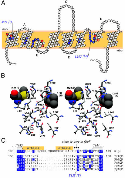
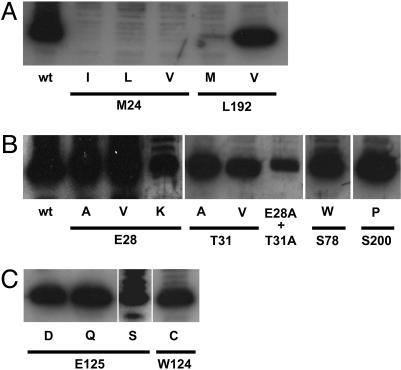
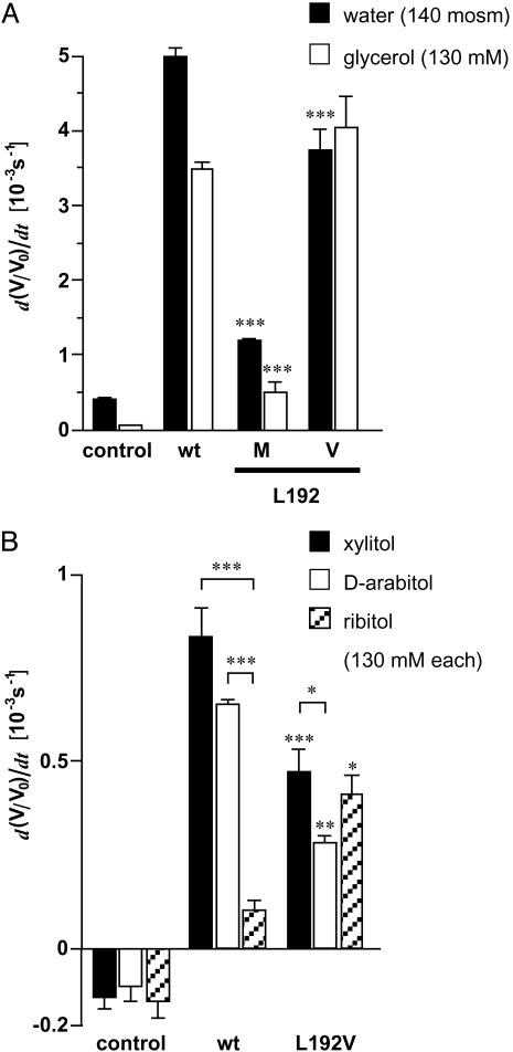
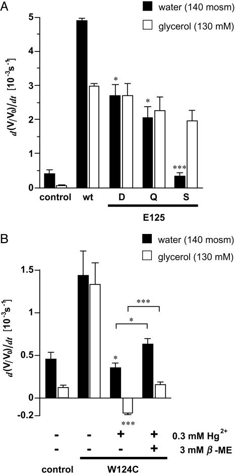
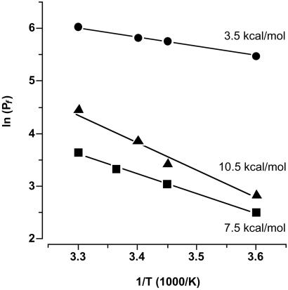
The selectivity of aquaporins for water and solutes is determined by pore diameter. Paradoxically, the wider pores of glycerol facilitators restrict water passage by an unknown mechanism. Earlier we characterized an aquaglyceroporin from Plasmodium falciparum with high permeability for both glycerol and water. We use point mutations to demonstrate that amino acids directly lining the pore are not responsible for the excellent water permeability of the Plasmodium aquaglyceroporin but affect permeability of pentitols. Within a conserved WET triad in the extracellular C-loop we identified a Plasmodium aquaglyceroporin-specific glutamate (E125) located in proximity to a conserved arginine (R196) at the pore mouth. Mutation of E125 to serine largely abolished water permeability. Concomitantly, the activation energy for water permeation was increased by 4 kcal/mol. Mutation of the adjacent tryptophan to cysteine led to irreversible inhibition of water passage by Hg(2+). This unequivocally proves the proximity of the couple W124/E125 close to the pore mouth. We conclude that in the Plasmodium aquaglyceroporin the electrostatic environment at the extracellular pore entry regulates water permeability.


Figures

Fig. 1.
Fig. 1.
Sequence and structure comparisons of the aquaglyceroporins of Plasmodium and E. coli. (A) Topology prediction for PfAQP with the pore forming residues shaded blue. Compared to GlpF, only two residues differ in PfAQP within the selective part of the pore, namely M24(I22) and L192(M202). Red-shaded residues indicate six further sequence differences between PfAQP and GlpF in the immediate pore vicinity. Intra- and extracellular loops are labeled alphabetically. (B) Stereoview of the pore-lining amino acids (see blue-shaded residues in A) as predicted for PfAQP from a sequence projection on the GlpF structure. The two differences in this area [M24(I22) and L192(M202)] are drawn in space filling mode. Bars on the right indicate the proposed filter regions of the aquaporins, i.e., the upper aromatic/arginine filter [W50(48), F190(200), and R196(206)] and the two asparagines in the pore center [N70(68) and N193(203)]. The color scheme follows convention with carbon in gray, nitrogen in blue, oxygen in red, and sulfur in yellow shading. (C) Alignment of C-loops from GlpF and aquaglyceroporins from P. falciparum (PfAQP), P. berghei (PbAQP), P. chabaudi (PcAQP), P. knowlesi (PkAQP), and P. yoelii (PyAQP). Matches between GlpF and the Plasmodium aquaglyceroporins are shaded in blue. The superscript dots mark the conserved amino acid triad, which is fixed at the pore entry in GlpF. The Plasmodium-specific E125(S136) is highlighted below. Boxes denote the helical regions in the C-loop of GlpF.
Fig. 2.
Fig. 2.
Expression of PfAQP constructs by Western blotting with Xenopus oocyte membranes and an antiserum specific to the C terminus of PfAQP. (A) Mutations in the pore lining. (B) Mutations in the pore vicinity. (C) Mutations in the C-loop. Band broadening and multiple bands may be caused by incomplete secondary modifications and/or unspecific labeling.
Fig. 3.
Fig. 3.
Swelling of oocytes expressing PfAQP constructs with mutations in the pore lining. (A) Swelling rates for water and glycerol (n = 4–6). (B) Permeability of pentitols through PfAQP and the L192V mutant (n = 3–5). Asterisks above the error bars indicate significant differences in the flux rates of the same solute between wild-type and mutant PfAQP. Asterisks above brackets indicate significant differences in the flux rates of different solutes through the same channel protein (*, P < 0.05; **, P < 0.02; ***, P < 0.01).
Fig. 4.
Fig. 4.
Swelling rates of oocytes expressing PfAQP with C-loop mutations. (A) Water and glycerol swelling rates (n = 5–8 and 3–5 for water and glycerol, respectively). Glycerol permeability was not significantly different. Asterisks above the error bars indicate significant differences in the water permeability between wild-type and mutant PfAQP. (B) Effect of Hg2+ on water and glycerol permeation of PfAQP W124C. Swelling rates of oocytes were determined before and after 5 min incubation in ND96 medium with 0.3 mM Hg2+. Furthermore, swelling rates of oocytes are shown that were held in ND96 with 3 mM 2-mercaptoethanol for >10 min after the mercury inhibition (n = 3–6). Asterisks above the error bars indicate significant differences in the water and glycerol permeabilty before and after mercury treatment. Asterisks above brackets indicate significance levels of the recovery of water and glycerol permeability after incubation in 2-mercaptoethanol (*, P < 0.05; **, P < 0.02; ***, P < 0.01).
Fig. 5.
Fig. 5.
Arrhenius plot of the water permeability (Pf) through PfAQP wild-type (circles), PfAQP E125S (squares), and control oocyte membranes (triangles). Plotted are logarithms of the Pf values against reciprocals of the temperature at which the measurements were carried out (4–30°C). Activation energies were calculated from the slopes of the linear fits (n = 5).

References

    1. Borgnia, M., Nielsen, S., Engel, A. & Agre, P. (1999) Annu. Rev. Biochem. 68, 425–458. - PubMed
    1. Walz, T., Hirai, T., Murata, K., Heymann, J. B., Mitsuoka, K., Fujiyoshi, Y., Smith, B. L., Agre, P. & Engel, A. (1997) Nature 387, 624–627. - PubMed
    1. Cheng, A., van Hoek, A. N., Yeager, M., Verkman, A. S. & Mitra, A. K. (1997) Nature 387, 627–630. - PubMed
    1. Sui, H., Han, B. G., Lee, J. K., Walian, P. & Jap, B. K. (2001) Nature 414, 872–878. - PubMed
    1. Braun, T., Philippsen, A., Wirtz, S., Borgnia, M. J., Agre, P., Kühlbrandt, W., Engel, A. & Stahlberg, H. (2000) EMBO Rep. 1, 183–189. - PMC - PubMed

Publication types

LinkOut - more resources