. 2004 Jan 20;32(2):407-14.
doi: 10.1093/nar/gkh209. Print 2004.

RNase H2 of Saccharomyces cerevisiae is a complex of three proteins

Affiliations

RNase H2 of Saccharomyces cerevisiae is a complex of three proteins

Ho-Sang Jeong et al. Nucleic Acids Res. .

Erratum in

  • Nucleic Acids Res. 2004 Feb 24;32(4):1616

Abstract

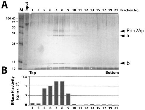
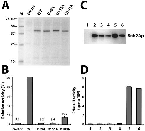
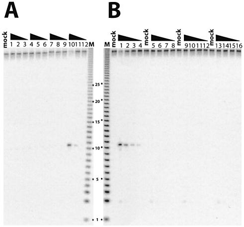
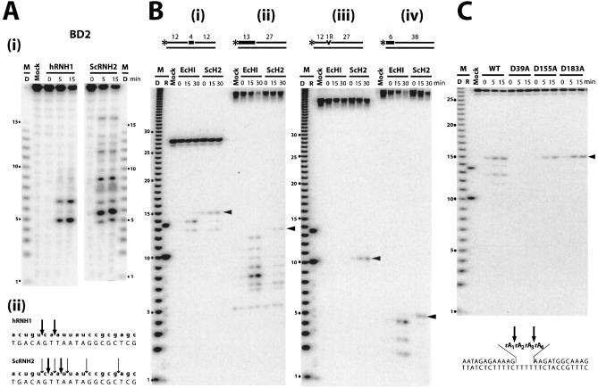
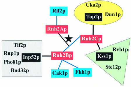
The composition of RNase H2 has been a long-standing problem. Whereas bacterial and archaeal RNases H2 are active as single polypeptides, the Saccharomyces cerevisiae homolog, Rnh2Ap, when expressed in Escherichia coli, fails to produce an active RNase H2. By affinity chromatography purification and identification of polypeptides associated with a tagged S.cerevisiae Rnh2Ap, we obtained a complex of three proteins [Rnh2Ap (Rnh201p), Ydr279p (Rnh202p) and Ylr154p (Rnh203p)] that together are necessary and sufficient for RNase H2 activity [correction]. Deletion of the gene encoding any one of the proteins or mutations in the catalytic site in Rnh2A led to loss of RNase H2 activity. Even when S.cerevisiae RNase H2 is catalytically compromised, it still exhibits a preference for cleavage of the phosphodiester bond on the 5' side of a ribonucleotide-deoxyribonucleotide sequence in substrates mimicking RNA-primed Okazaki fragments or a single ribonucleotide embedded in a duplex DNA. Interestingly, Ydr279p and Ylr154p have homologous proteins only in closely related species. The multisubunit nature of S.cerevisiae RNase H2 may be important both for structural purposes and to provide a means of interacting with other proteins involved in DNA replication/repair and transcription.


Figures

Figure 1
Figure 1
Purification of S.cerevisiae RNase H2 by glycerol gradient ultracentrifugation following FLAG immunopurification. (A) A low copy plasmid carrying the tagged RNH2A gene driven by the RNH2A promoter was used to prepare extracts. Aliquots of glycerol gradient fractions were analyzed by silver staining after SDS–PAGE. Rnh2Ap (marked by an arrow) with two other polypetides (a and b). In several preparations, additional bands were identified as Ydr279p and Ylr154p by mass spectrometry of tryptic peptides (data not shown). (B) RNase H assays of the glycerol gradient. A 1 µl aliquot of each fraction was assayed for RNase H activity using 32P-labeled poly(rA)–poly(dT) as substrate.
Figure 2
Figure 2
Requirement for Rnh2Ap and two additional polypeptides for RNase H2 activity. (A) Silver-stained SDS–PAGE of samples. FLAG- immunopurified Asp→Ala mutant proteins of Rnh2Ap from S.cerevisiae expressed from a multicopy plasmid were analyzed by SDS–PAGE. (B) RNase H assay of Asp→Ala substitution mutants. Similar amounts of Rnh2Ap were used for assays based on quantification of total protein. Activity is from the mean of three replicate assays and is expressed as a percentage of the values for the wild-type enzyme. Error bars represent standard deviation. (C) Western blot analysis of purified recombinant Rnh2Ap in deletion strains: ydr279wΔ (lane 2), ylr154cΔ (lane 3) ydr279wΔ ylr154CΔ (lane 4), rnh2Δ (lane 5) and wild type (lane 6). Lane 1 is a control assay with an equivalent volume of an extract from the ydr279wΔ strain containing the vector. Anti-HA antibody was used as probe for western blotting. (D) RNase H assays of partially purified RNase H2 from various deletion strains. The amounts of Rnh2Ap used for assays were adjusted based on western blotting intensity. Lane numbers correspond to those of (C), and the values represent the mean of three assays.
Figure 3
Figure 3
RNase H assays using monoribonucleotide-embedded double-stranded DNA as substrate. DNA12–RNA1–DNA27/DNA40 was used as substrate and similar amounts of protein (determined by protein assays) were assayed. The largest amount of protein assayed was 10 times that used in the adjacent lane, and so forth. The triangle indicates decreasing concentrations. Mock is with no enzyme added. (A) His tag affinity purification of E.coli expressed Rnh2Ap (lanes 1–3), Ydr279p (lanes 4–6), Ylr154p (lanes 7–9) and all three polypeptides expressed together (lanes 10–12). (B) Crude extracts of S.cerevisiae from wild-type (lanes 1–4), rnh2AΔ (lanes 5–8), ydr279wΔ (lanes 9–12) and ylr154cΔ (lanes 13–16).
Figure 4
Figure 4
Comparison of cleavage patterns of RNase H2 and RNases HI/H1. Digests were for the times (min) indicated above each lane. Molecular size markers are indicated as D [products of digestion of 32P-labeled poly(rA)–poly(dT) by E.coli RNase HI] and R synthetic oligoribonucleotide markers of 10 and 13 nucleotides. Arrowheads mark the cleavages leaving a single ribonucleotide attached to the downstream DNA. (A) (i) Digestion of BD2 substrate by human RNase H1 and S.cerevisiae RNase H2 analyzed by 12% TBE–urea PAGE. (ii) Sites of cleavage are marked by arrows, with the relative frequency indicated by the size of the arrow. (B) Comparison of digestion patterns generated by E.coli RNase HI and S.cerevisiae RNase H2 on (i) DNA12–RNA4–DNA12/DNA28; (ii) RNA13–DNA27/DNA40; (iii) DNA12–RNA1–DNA27/DNA40; and (iv) RNA6–DNA38/DNA40 as substrates. (C) Digestion of DNA12–RNA4–DNA12/DNA28 as substrate by S.cerevisiae RNase H2 and the three D→A mutant proteins (D39A, D155A and D183A). The four internal rA residues are shown as rA1rA2rA3rA4.
Figure 5
Figure 5
Interactions of RNase H2 subunits. The three subunits of RNase H2 are indicated in red and connected by a star. Interactions obtained using the yeast two-hybrid system are indicated by blue connecting lines; note that Rnh2B and Rnh2C are connected by blue lines. Complexes in which Rnh2B and Rnh2C were found by mass spectrometric analysis are shown in groups by color. The proteins used to isolate the complexes are indicated in black and connected by lines to the RNase H2 subunits. The Top2p complex is in the yellow oval, the Kss1p complex is in the green triangle, and the Inp52p complex is in the gray square.

References

    1. Crouch R.J. and Cerritelli,S.M. (1998) RNases H of S.cerevisiae, S.pombe, C.fasciculata, and N.crassa. In Crouch,R.J. and Toulmé,J.J. (eds), Ribonucleases H. INSERM, Paris, pp. 79–100.
    1. Ohtani N., Haruki,M., Morikawa,M., Crouch,R.J., Itaya,M. and Kanaya,S. (1999) Identification of the genes encoding Mn2+-dependent RNase I–III and Mg2+-dependent RNase HIII from Bacillus subtilis: classification of RNases H into three families. Biochemistry, 38, 605–618. - PubMed
    1. Muroya A., Tsuchiya,D., Ishikawa,M., Haruki,M., Morikawa,M., Kanaya,S. and Morikawa,K. (2001) Catalytic center of an archaeal type 2 ribonuclease H as revealed by X-ray crystallographic and mutational analyses. Protein Sci., 10, 707–714. - PMC - PubMed
    1. Chapados B.R., Chai,Q., Hosfield,D.J., Qiu,J.Z., Shen,B.H. and Tainer,J.A. (2001) Structural biochemistry of a type 2 RNase H: RNA primer recognition and removal during DNA replication. J. Mol. Biol., 307, 541–556. - PubMed
    1. Ohtani N., Haruki,M., Morikawa,M. and Kanaya,S. (1999) Molecular diversities of RNases H. J. Biosci. Bioeng., 88, 12–19. - PubMed

MeSH terms