. 2004 Jan;2(1):E2.
doi: 10.1371/journal.pbio.0020002. Epub 2003 Nov 24.

JAMM: a metalloprotease-like zinc site in the proteasome and signalosome

Affiliations

JAMM: a metalloprotease-like zinc site in the proteasome and signalosome

Xavier I Ambroggio et al. PLoS Biol. 2004 Jan.

Abstract

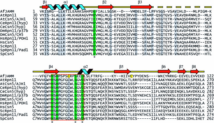
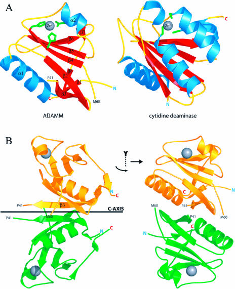
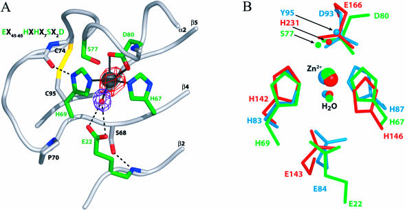
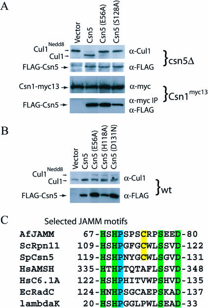
The JAMM (JAB1/MPN/Mov34 metalloenzyme) motif in Rpn11 and Csn5 underlies isopeptidase activities intrinsic to the proteasome and signalosome, respectively. We show here that the archaebacterial protein AfJAMM possesses the key features of a zinc metalloprotease, yet with a distinct fold. The histidine and aspartic acid of the conserved EX(n)HS/THX(7)SXXD motif coordinate a zinc, whereas the glutamic acid hydrogen-bonds an aqua ligand. By analogy to the active site of thermolysin, we predict that the glutamic acid serves as an acid-base catalyst and the second serine stabilizes a tetrahedral intermediate. Mutagenesis of Csn5 confirms these residues are required for Nedd8 isopeptidase activity. The active site-like architecture specified by the JAMM motif motivates structure-based approaches to the study of JAMM domain proteins and the development of therapeutic proteasome and signalosome inhibitors.


Conflict of interest statement

The authors have declared that no conflicts of interest exist.

Figures

Figure 1
Figure 1. Alignment of Eukaryotic JAMM Domains with AfJAMM
Eukaryotic JAMM domain proteins were aligned with AfJAMM using ClustalX and manually refined. Sequences are named with a two-letter code corresponding to the genus and species of the respective organism followed by the name of the protein (see Supporting Information for accession numbers), and ‘hyp’ is an abbreviation for hypothetical. The JAMM motif comprises the residues highlighted in green (E22, H67, H69, S77, and D80), and the active site core is surrounded by a red box. Conserved residues are highlighted in gray. The disulfide cysteine residues are highlighted in yellow (C74, C95). Active site residues that were mutated in S. pombe Csn5 are marked with an asterisk beneath the alignment. The secondary structure of AfJAMM is indicated above the sequence; helices are blue, sheets are red arrows, and loops are yellow lines. The dashed yellow line indicates a loop (F42–G58) that is disordered in the crystal.
Figure 2
Figure 2. Crystal Structure of AfJAMM
(A) On the left, the AfJAMM protomer is presented. The amino and carboxyl termini are marked by N and C. The catalytic zinc atom is depicted as a gray sphere. The zinc ligands (H67, H69, and D80) are colored in green. Secondary structure elements are numbered α1–α2 and β1–β8. The amino acids that mark the beginning and end of the disordered loop (P41–M60) are labeled. On the right, the crystal structure of the cytidine deaminase protomer is shown in the same orientation as AfJAMM to highlight the fold likeness as well as the similarly situated zinc-binding sites. The zinc ligands (C53, C86, and C89) are colored in green. (B) The dimer in the asymmetric unit of AfJAMM crystals. The side view is obtained by rotating the monomer in (A) by 90° as indicated by the quarter-arrow around the y-axis. The gold protomer is related to the green protomer by a 180° rotation around the crystallographic c-axis (shown as a black bar in the side view) and a translation of 3.38 Å.
Figure 3
Figure 3. Metalloprotease-Like Active Site of AfJAMM
(A) The active site of AfJAMM is shown centered around the catalytic zinc ion, which is represented as a dark gray sphere surrounded by anomalous cross Fourier difference density (contoured at 9.5 σ) colored in red. The aqua ligand, which lies at 2.9 Å from the zinc, is shown as a red sphere surrounded by purple density (contoured at 3 σ) of an Fobs – Fcalc map, in which the aqua ligand was omitted from the calculation. Residues that underlie isopeptide bond cleavage are shown in green. The carboxylate oxygen atoms of D80 lie 2.2 Å from the zinc. The Nɛ2 atoms of H67 and H69 lie 2.1 Å from the zinc. The carboxylate oxygen atoms of E22 lie 3.2–3.5 Å from the aqua ligand and 4.5–5.0 Å from the zinc. Ancillary active site residues and the backbone (ribbon diagram) are shown in grey. The disulfide bond that links C74 to C95 is shown in yellow. The JAMM motif is shown in the upper lefthand corner for reference. (B) Superimposition of active site residues in ScNP, thermolysin, and AfJAMM. AfJAMM is in green, ScNP in blue, and thermolysin in red. For clarity only, the sidechains from the residues that bind the zinc or aqua ligands are shown in their entirety. In addition, atoms that stabilize the putative tetrahedral intermediate are shown. These include Oγ of S77 in AfJAMM, Oη of Y95 in ScNP, and the Nɛ2 of H231 in thermolysin.
Figure 4
Figure 4. Mutations in the JAMM Motif of Csn5 Abrogate the Deneddylating Activity of the CSN
(A) Mutations in the glutamic acid (E56A) that positions the aqua ligand and in the proposed catalytic serine (S128A) of Csn5 disrupt deneddylation of Cul1 by CSN but have no effect on assembly with Csn1. A csn5Δ strain of S. pombe was transformed with an empty pREP-41 plasmid (lane 1) or with the plasmid encoding FLAG tagged: Csn5 (lane 2), Csn5E56A (lane 3), or Csn5S128A (lane 4). Whole-cell lysates were used for Western blot analysis with anti-Cul1 antibodies (top gel) and anti-FLAG antibodies (second from top). A strain with a myc13-tagged Csn1 was transformed with the above plasmids, and whole-cell lysates were used for Western blot analysis. Antibodies to the Myc tag were used to detect Csn1myc13 (third from top), and were used to pull down Csn1myc13 and subsequently blot with anti-FLAG antibodies to detect coprecipitated Csn5 mutant proteins (bottom gel). (B) Mutations in the JAMM motif display a modest dominant-negative phenotype. Western blot analysis of crude cell lysates was performed as described in (A). (C) Selected JAMM motifs from proteins of diverse functions. The canonical JAMM motif residues are highlighted in green. The conserved proline is highlighted in blue, and semiconserved cysteine is highlighted in yellow.

References

    1. Adams J. Development of the proteasome inhibitor PS-341. Oncologist. 2002;7:9–16. - PubMed
    1. Aravind L, Ponting CP. Homologues of 26S proteasome subunits are regulators of transcription and translation. Protein Sci. 1998;7:1250–1254. - PMC - PubMed
    1. Auld DS. Removal and replacement of metal-ions in metallopeptidases. Methods Enzymol. 1995;248:228–242. - PubMed
    1. Bartlett PA, Marlowe CK. Possible role for water dissociation in the slow binding of phosphorus-containing transition-state-analogue inhibitors of thermolysin. Biochemistry. 1987;26:8553–8561. - PubMed
    1. Brown CK, Madauss K, Lian W, Beck MR, Tolbert WD, et al. Structure of neurolysin reveals a deep channel that limits substrate access. Proc Natl Acad Sci U S A. 2001;98:3127–3132. - PMC - PubMed

Publication types

MeSH terms

Associated data