. 2004 Jan;2(1):E3.
doi: 10.1371/journal.pbio.0020003. Epub 2004 Jan 20.

Dorsoventral patterning of the mouse coat by Tbx15

Affiliations

Dorsoventral patterning of the mouse coat by Tbx15

Sophie I Candille et al. PLoS Biol. 2004 Jan.

Abstract

Many members of the animal kingdom display coat or skin color differences along their dorsoventral axis. To determine the mechanisms that control regional differences in pigmentation, we have studied how a classical mouse mutation, droopy ear (de(H)), affects dorsoventral skin characteristics, especially those under control of the Agouti gene. Mice carrying the Agouti allele black-and-tan (a(t)) normally have a sharp boundary between dorsal black hair and yellow ventral hair; the de(H) mutation raises the pigmentation boundary, producing an apparent dorsal-to-ventral transformation. We identify a 216 kb deletion in de(H) that removes all but the first exon of the Tbx15 gene, whose embryonic expression in developing mesenchyme correlates with pigmentary and skeletal malformations observed in de(H)/de(H) animals. Construction of a targeted allele of Tbx15 confirmed that the de(H) phenotype was caused by Tbx15 loss of function. Early embryonic expression of Tbx15 in dorsal mesenchyme is complementary to Agouti expression in ventral mesenchyme; in the absence of Tbx15, expression of Agouti in both embryos and postnatal animals is displaced dorsally. Transplantation experiments demonstrate that positional identity of the skin with regard to dorsoventral pigmentation differences is acquired by E12.5, which is shortly after early embryonic expression of Tbx15. Fate-mapping studies show that the dorsoventral pigmentation boundary is not in register with a previously identified dermal cell lineage boundary, but rather with the limb dorsoventral boundary. Embryonic expression of Tbx15 in dorsolateral mesenchyme provides an instructional cue required to establish the future positional identity of dorsal dermis. These findings represent a novel role for T-box gene action in embryonic development, identify a previously unappreciated aspect of dorsoventral patterning that is widely represented in furred mammals, and provide insight into the mechanisms that underlie region-specific differences in body morphology.


Conflict of interest statement

The authors have declared that no conflicts of interest exist.

Figures

Figure 1
Figure 1. Dorsoventral Skin Characteristics
(A) Skin slices from animals of different age and genotype demonstrate similar patterns of hair-length variation along the dorsoventral axis (scale bar = 1 cm). (B) Enlarged area from (A), demonstrating the transition in hair length and color in at/at mice (scale bar = 0.375 cm). (C) Proportional hair length for (A) plotted as a function of relative position along the dorsoventral axis. (D) Hair length plotted as a function of absolute position along the dorsoventral axis for 8-wk-old BA strain mice. (E) Proportion of zigzag hairs (± SEM) differs slightly between dorsum and ventrum of inbred mice (p < 0.0001, χ2 test, n = 1,958, 1,477, 1,579, 1,502). (F) Differences in dorsal and ventral skin development at P4.5 (scale bar = 1 mm, upper; 200 μm, lower). (G) Differences in hair melanin content and DOPA staining for dorsum (d), flank (f), and ventrum (v) in ae/ae and at/at mice. The upper panel also demonstrates a cream-colored appearance of the at/at ventrum. The middle panel shows representative awls (scale bar = 100 μm). The lower panel shows DOPA-stained dermis (scale bar = 200 μm).
Figure 2
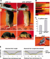
Figure 2. The deH Pigmentation Phenotype
(A) 10-wk-old deH/deH and nonmutant animals on a at background. A thin stripe of yellow hair normally separates the dorsal black hairs from the ventral cream hairs. In deH, the yellow stripe is extended dorsally, and the boundary between the yellow and the black hairs is fuzzier. (B) Skin slices taken from 1.5-mo-old deH/deH and nonmutant littermates (scale bar = 0.5 cm). (C) Proportion of total skin area as determined by observation of pelts taken from the interlimb region. The proportion occupied by the yellow lateral compartment (± SEM) differs between mutant and nonmutant littermate flanks (p < 0.0005, paired t-test, n = 6 pairs). There is also (data not shown) a small increase in the proportion of total skin area occupied by the ventral cream-colored compartment, 47.9 % in mutant compared to 37.8% in nonmutant (p < 0.005, paired t-test, n = 6 pairs). (D) On an ae/ae background, the extent of dorsal skin pigmentation is reduced in deH/deH neonates (P3.5). (E) Hair length in a representative pair of 1.5-mo-old deH/deH and nonmutant littermates, averaged over three skin slices at different rostrocaudal levels, and plotted as a function of the absolute distance from middorsum or the percentage of total slice length.
Figure 3
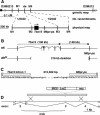
Figure 3. Molecular Genetics of deH and Tbx15
(A) Genetic and physical map, as described in the text. Markers M1 to M3 are SSCP markers generated from a BAC contig of the region; marker M4 is STS 16.MMHAP32FLF1 and was also used as an SSCP marker. M2 and M3, which flank the Tbx15 and M6pr-ps on the UCSC genome browser map and lie 634 kb apart, were nonrecombinant with deH in 2340 meioses. (B) The deH mutation is a deletion that starts in Tbx15 intron 1 and ends in the M6pr-ps. (C) Sequence of deletion breakpoints. (D) Diagram of Tbx15LacZ allele constructed by gene targeting. As described in the text, this allele is predicted to give rise to a protein truncated after approximately 154 codons and is lacking critical residues of the T box. Heterozygotes for the targeted allele exhibit normal size, morphology, and hair-color patterns, but homozygotes and Tbx15LacZ/deH compound heterozygotes are identical to deH homozygotes.
Figure 4
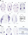
Figure 4. Developmental Expression of Tbx15
(A) At E12.5, transverse sections at different levels show expression in head mesenchyme (a and b); myotome, occipital, and periocular mesenchyme (b); palatal shelf, cervical sclerotome, and nasal cartilage (c); maxillary and mandibular processes (d); limbs (e); and myotome and lateral mesenchyme (e and f) (scale bars = 500 μm). (B) Transverse sections through the flank at different times show expression in lateral mesenchyme (E11.5), expanding dorsally at E12.5, and both ventrally and dorsally at E13.5, detectable in loose mesenchyme underlying the dermis and the abdominal and subcutaneous muscles (scale bar = 500 μm). At P3.5, Tbx15 is expressed in the entire dermis and is most strongly expressed in dermal sheaths (scale bar = 200 μm).
Figure 5
Figure 5. Embryonic Expression of Tbx15 Compared to Agouti in at/at Mice
(A and C) Tbx15. (B and D) Agouti. At E12.5, expression of Tbx15 in dorsal skin is approximately complementary to that of Agouti in ventral skin. At E14.5, the levels of expression for both genes are lower, but Tbx15 expression has expanded ventrally and overlaps extensively with that of Agouti. In all four panels, arrows mark the approximate ventral limit of Tbx15 and the approximate dorsal limit of Agouti (scale bars = 500 μm).
Figure 6
Figure 6. Effect of deH on Agouti Expression
Comparable sections from at/at; deH/deH and at/at; +/+ littermates. (A) At E14.5, deH/deH embryos have a smaller body cavity and loose skin within which Agouti expression appears to be shifted dorsally, as marked by arrows (scale bars = 500 μm). (B) At P4.5, Agouti expression in both dorsal and ventral skin is similar in deH/deH compared to nonmutant, but in the midflank region, there is increased Agouti expression in deH/deH, especially in the upper dermis (scale bars = 200 μm). Sections shown are representative of two mutant and two nonmutant samples examined at each time.
Figure 7
Figure 7. Embryonic Establishment of Dorsoventral Skin Patterning
Pieces of skin from dorsal, flank, and ventral regions of at/a E12.5 embryos were transplanted into the testes of congenic animals as described in the text. Hair color of the grafts was examined 3 wk later. Grafts of ventral embryonic skin (n = 3) produced yellow hairs, dorsal embryonic skin (n = 4) produced black hairs, and flank embryonic skin produced mostly (13 out of 15) black and yellow hairs in distinct regions as shown. In parallel, in situ hybridization studies revealed that the embryonic flank contains the boundary of expression between Agouti and Tbx15 (scale bars = 1 mm for hairs and 200 μm for in situ hybridization results).
Figure 8
Figure 8. Comparison of the Dorsoventral at/at Pigmentation Boundary to the Lateral Somitic Frontier
(A) Dorsoventral slices of skin from at the midtrunk region prepared such that the dorsal midline lies in the center of the slice. Sections were taken at P1.5 (a) or P4.5 (b–e) from at/at or R26R/+; Tg.Hoxb6-Cre/+ mice (the latter were stained with X-Gal), as described in Materials and Methods. For purposes of comparison, images were proportionally scaled. The boundary of X-Gal staining marks dermis derived from lateral plate versus dermis derived from mesoderm (the lateral somitic frontier) and lies more dorsal than the at/at pigmentation boundary. (B) Quantitation of mean (± SEM) dorsal pigmentation area (n = 5) and somite-derived dermis area (n = 3) shows a significant difference (p < 0.005, t-test). (C) RNA in situ hybridization showing that Tbx15 expression at E11.5 is complementary to En1 expression on the flank (scale bars = 200 μm). The arrow indicates the boundary between the expression domains of the two genes.
Figure 9
Figure 9. Model for Acquisition of Dorsoventral Patterning in the Trunk and the Role of Tbx15
(A) A tricolor pigmentation pattern is generated by the combination of distinct mechanisms that affect distribution of Agouti mRNA and histochemical staining for melanocytes; effects of the latter mechanism by itself are evident in ae/ae mice (see Figure 1). In at/at mice, reduced hair melanocyte activity and high levels of Agouti mRNA in the ventrum lead to a cream color; as melanocyte activity gradually increases towards the dorsum, a lateral stripe is apparent on the flank. The distributions of Agouti mRNA and histochemical staining for melanocytes are both affected by Tbx15 and are externally evident by a widening of the lateral stripe and an increased proportion of total skin occupied by the cream-colored area. (B) The lateral yellow stripe in at/at mice lies at the same level as the limb dorsoventral boundary. As described in the text, we propose that distinct dorsoventral compartments in ectoderm of the trunk provide an instructional cue to the mesoderm, leading to expression of Tbx15 in dorsal trunk mesenchyme and acquisition of dorsal dermis character. In the absence of Tbx15, dorsal mesenchyme assumes ventral characteristics instead.

References

    1. Agulnik SI, Papaioannou VE, Silver LM. Cloning, mapping, and expression analysis of TBX15, a new member of the T-box gene family. Genomics. 1998;51:68–75. - PubMed
    1. Altabef M, Clarke JD, Tickle C. Dorso-ventral ectodermal compartments and origin of apical ectodermal ridge in developing chick limb. Development. 1997;124:4547–4556. - PubMed
    1. Altabef M, Logan C, Tickle C, Lumsden A. Engrailed-1 misexpression in chick embryos prevents apical ridge formation but preserves segregation of dorsal and ventral ectodermal compartments. Dev Biol. 2000;222:307–316. - PubMed
    1. Barsh G, Gunn T, He L, Schlossman S, Duke-Cohan J. Biochemical and genetic studies of pigment-type switching. Pigment Cell Res. 2000;13(Suppl 8):48–53. - PubMed
    1. Begemann G, Gibert Y, Meyer A, Ingham PW. Cloning of zebrafish T-box genes tbx15 and tbx18 and their expression during embryonic development. Mech Dev. 2002;114:137–141. - PubMed

Publication types

MeSH terms

Substances

LinkOut - more resources