. 2004 Jan;2(1):E4.
doi: 10.1371/journal.pbio.0020004. Epub 2004 Jan 20.

Cell-passage activity is required for the malarial parasite to cross the liver sinusoidal cell layer

Affiliations

Cell-passage activity is required for the malarial parasite to cross the liver sinusoidal cell layer

Tomoko Ishino et al. PLoS Biol. 2004 Jan.

Abstract

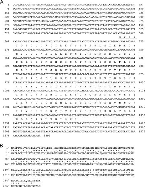
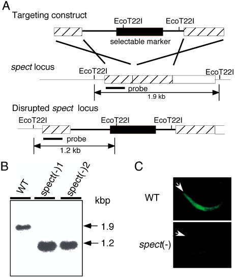
Liver infection is an obligatory step in malarial transmission, but it remains unclear how the sporozoites gain access to the hepatocytes, which are separated from the circulatory system by the liver sinusoidal cell layer. We found that a novel microneme protein, named sporozoite microneme protein essential for cell traversal (SPECT), is produced by the liver-infective sporozoite of the rodent malaria parasite, Plasmodium berghei. Targeted disruption of the spect gene greatly reduced sporozoite infectivity to the liver. In vitro cell invasion assays revealed that these disruptants can infect hepatocytes normally but completely lack their cell passage ability. Their apparent liver infectivity was, however, restored by depletion of Kupffer cells, hepatic macrophages included in the sinusoidal cell layer. These results show that malarial sporozoites access hepatocytes through the liver sinusoidal cell layer by cell traversal motility mediated by SPECT and strongly suggest that Kupffer cells are main routes for this passage. Our findings may open the way for novel malaria transmission-blocking strategies that target molecules involved in sporozoite migration to the hepatocyte.


Conflict of interest statement

The authors have declared that no conflicts of interest exist.

Figures

Figure 1
Figure 1. Sequence Analysis of spect cDNA
(A) Nucleotide sequence of spect cDNA (top lane) and the deduced amino acid sequence (bottom lane) are shown. The predicted N-terminal signal sequence is underlined. The numbers indicate positions of the nucleotides starting from the 5′ end. The asterisks indicate the termination codon. (B) A comparison of deduced amino acid sequences of P. berghei spect (top) and P. falciparum spect (bottom). Gaps are introduced to obtain optical matching by using GENETIX-MAC software. Asterisks or dots show conserved or similar residues, respectively. The amino acid numbers from the first Met residue are shown on the left of each line.
Figure 2
Figure 2. SPECT Is a Microneme Protein Specifically Produced in the Liver-Infective Sporozoite Stage
(A) Indirect immunofluorescence microscopy of all four invasive forms of the malarial parasite (indicated over the panel). Parasites were stained with primary antibodies against SPECT, followed by FITC-conjugated secondary antibodies. SPECT was detected only in the salivary gland sporozoite, the liver-infective stage. The corresponding phase-contrast or DAPI-stained image (Phase or DAPI) is shown under each image. Scale bars, 5 μm (B) Western blot analysis of SPECT production in the midgut sporozoite (M) and the salivary gland sporozoite (S). Lysate of 500,000 sporozoites was loaded onto each lane and detected with the same antibody used in (A). SPECT was detected as a single band of 22 kDa (arrowhead) only in the salivary gland sporozoite. (C) Immunoelectron microscopy of sporozoites in the salivary gland. Ultrathin sections of a mosquito salivary gland infected with sporozoites were incubated with the same antibody used in (A) followed by secondary antibodies conjugated with gold particles (15 nm). Particles were localized to micronemes (Mn) but not to rhoptories (Rh). Axial (inset) and vertical images are shown. Scale bars, 0.5 μm.
Figure 3
Figure 3. Targeted Disruption of the spect Gene
(A) Schematic representation of targeted disruption of the spect gene. The targeting vector (top) containing a selectable marker gene is integrated into the spect gene locus (middle) by double crossover. This recombination event resulted in the disruption of the spect gene and confers pyrimethamine resistance to disruptants (bottom). (B) Genomic Southern blot hybridization of wild-type (WT) and spect(−) populations. Genomic DNA isolated from the respective parasite populations was digested with EcoT22I and hybridized with the probe indicated in (A) by a solid bar. By integration of the targeting construct, the size of detected fragments was decreased from 1.9 kbp to 1.2 kbp. The result is shown for two independently prepared populations, spect(−)1 and spect(−)2. (C) Immunofluorescence microscopy of the wild-type (WT) and spect(−) parasite. Sporozoites were collected from the salivary gland and stained with primary antibody against SPECT followed by FITC-conjugated secondary antibodies. The apical end of each sporozoite is indicated by an arrowhead.
Figure 4
Figure 4. Targeted Disruption of spect Results in Reduction of Sporozoite Infectivity to the Liver
(A) The salivary gland sporozoites of each parasite population were injected intravenously into five rats. The parasitemia of each rat was checked by a Giemsa-stained blood smear after inoculation on the days indicated. The average parasitemia after inoculation of 30,000 sporozoites was significantly low in disruptant populations, whereas their growth rates in the blood were essentially the same as the wild-type. The numbers of parasites inoculated were as follows: 30,000 spect(−)1 (open circles), 30,000 spect(−)2 (open triangles), 30,000 wild-type (filled circles), and 3,000 wild-type (filled squares). Values shown represent the mean parasitemia (± SEM) of five rats. (B) The salivary gland sporozoites (500,000) of wild-type or spect-disrupted parasites were inoculated intravenously into 3-wk-old rats. After 24 h, the livers were fixed with paraformaldehyde and frozen. The number of EEFs on each cryostat sections was estimated by indirect immunofluorescence analysis using anti-CS antiserum. Values shown represent the mean number of EEFs per square millimeter (± SEM) of at least three rats.
Figure 5
Figure 5. spect Disruption Results in Loss of Cell-Passage Activity of the Sporozoite
(A) spect disruption does not affect sporozoite ability to infect hepatocytes. (Top panel) Comparison of EEF numbers between disruptants (spect(−)) and wild-type (WT) parasites. Salivary gland sporozoites were added to HepG2 cells and cultured for 48 h. EEFs formed were counted after immunofluorescence staining with an antiserum against CS protein. (Bottom panels) Representative fluorescence stained images. (B) Sporozoites lacking SPECT cannot traverse HeLa cells. (Top) Comparison of cell-passage activity between disruptants and wild-type parasites. Salivary gland sporozoites were added to HeLa cells and incubated for 1 h with FITC-conjugated dextran (1 mg/ml). Cell-passage activity was estimated by the number of cells wounded by sporozoite passage, which were identified by cytosolic labeling with FITC-conjugated dextran. (Bottom) Representative fluorescence stained images. All data are mean numbers of EEFs or FITC-positive cells in a one-fifth area of an 8-well chamber slide with standard errors for at least three independent experiments.
Figure 6
Figure 6. Restoration of spect(−) Sporozoite Infectivity in Kupffer Cell-Depleted Rats
(A) Liposome-encapsulated Cl2MDP (filled points) or PBS (open) was injected intravenously into rats. After 48 h, 30,000 sporozoites of spect(−)1 (circles), spect(−)2 (triangles), or wild-type (squares) populations were inoculated intravenously. Parasitemia of each rat was checked by Giemsa-stained blood smears after inoculation on the days indicated. Values shown represent the mean parasitemia (± SEM) of five rats. (B) Salivary gland sporozoites (500,000) of each parasite population were inoculated intravenously into Kupffer cell-depleted rats. After 24 h, the livers were fixed with paraformaldehyde and frozen. The number of EEFs on each cryostat section was estimated by indirect immunofluorescence analysis using anti-CS antiserum. Values shown represent the mean number of EEFs per square millimeter (± SEM) of at least three rats.
Figure 7
Figure 7. Schematic Representation of Sporozoite Migration to and Infection of Hepatocytes
(Left) Sporozoites migrate to the space of Disse through the Kupffer cells. [1] The sporozoite (Sp) in the circulatory system is sequestered to the sinusoidal endothelial cell (EC) by specific recognition of the cell surface or glycosaminoglycans extending from the hepatocytes (He) through fenestration. [2] The sporozoite begins to glide on the epithelial cell surface. [3] Encountering a Kupffer cell (KC), the sporozoite ruptures the plasma membrane, passes through the cell, and enters into the space of Disse. Thus, the sporozoite gains access to hepatocytes. This step requires SPECT. [4] The sporozoite infects a hepatocyte with formation of a vacuole and develops into EEF in the hepatocyte. (Right) An alternative route to the hepatocyte. A small number of sporozoites, which find gaps in the sinusoidal layer while gliding, migrate to hepatocytes directly through the openings without need for cell passage and infect the hepatocytes. Likewise, in Kupffer cell-depleted rats, both wild-type and spect(−) sporozoites can enter hepatocytes through numerous gaps present between the sinusoidal endothelial cells.

References

    1. Bouwens L, Baekeland M, De Zanger R, Wisse E. Quantitation, tissue distribution and proliferation kinetics of Kupffer cells in normal rat liver. Hepatology. 1986;6:718–722. - PubMed
    1. Carlton JM, Angiuoli SV, Suh BB, Kooij TW, Pertea M, et al. Genome sequence and comparative analysis of the model rodent malaria parasite Plasmodium yoelii yoelii . Nature. 2002;419:512–519. - PubMed
    1. Cerami C, Frevert U, Sinnis P, Takacs B, Clavijo P, et al. The basolateral domain of the hepatocyte plasma membrane bears receptors for the circumsporozoite protein of Plasmodium falciparum sporozoites. Cell. 1992;70:1021–1033. - PubMed
    1. Gardner MJ, Hall N, Fung E, White O, Berriman M, et al. Genome sequence of the human malaria parasite Plasmodium falciparum . Nature. 2002;419:498–511. - PMC - PubMed
    1. Hoffman SL, Franke ED, Hollingdale MR, Druihe P. Attacking the infected hepatocyte. In: Hoffman SL, editor. Malaria vaccine development. Washington, DC: American Society for Microbiology Press. pp. 1996:35–75.

Publication types

MeSH terms

Substances