. 2004 Jan;2(1):E6.
doi: 10.1371/journal.pbio.0020006. Epub 2004 Jan 20.

HIV-1 Nef binds the DOCK2-ELMO1 complex to activate rac and inhibit lymphocyte chemotaxis

Affiliations

HIV-1 Nef binds the DOCK2-ELMO1 complex to activate rac and inhibit lymphocyte chemotaxis

Ajit Janardhan et al. PLoS Biol. 2004 Jan.

Abstract

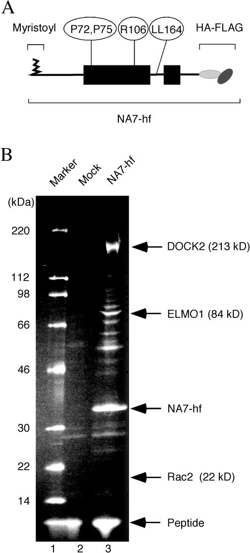
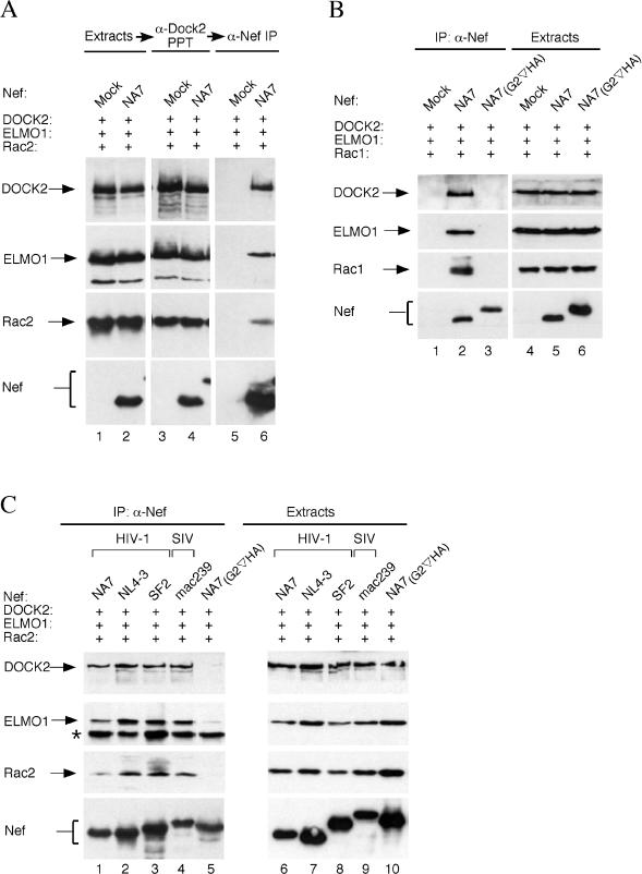
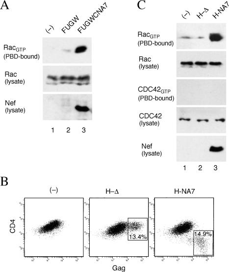
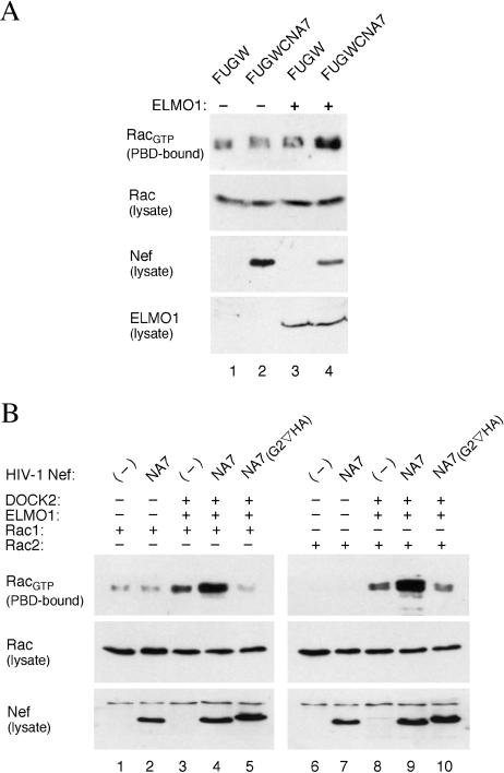
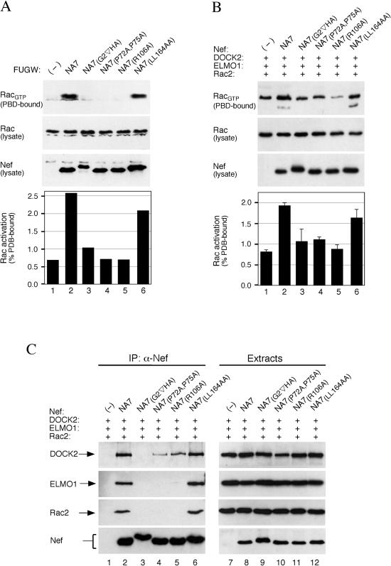
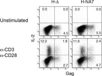
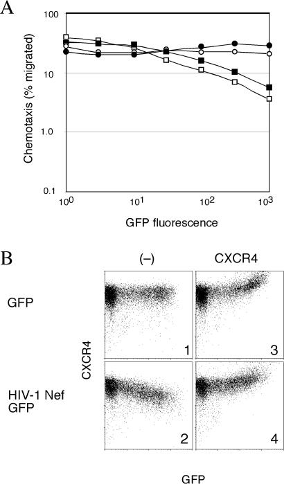
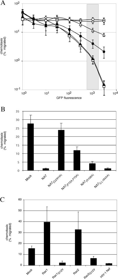
The infectious cycle of primate lentiviruses is intimately linked to interactions between cells of the immune system. Nef, a potent virulence factor, alters cellular environments to increase lentiviral replication in the host, yet the mechanisms underlying these effects have remained elusive. Since Nef likely functions as an adaptor protein, we exploited a proteomic approach to directly identify molecules that Nef targets to subvert the signaling machinery in T cells. We purified to near homogeneity a major Nef-associated protein complex from T cells and identified by mass spectroscopy its subunits as DOCK2-ELMO1, a key activator of Rac in antigen- and chemokine-initiated signaling pathways, and Rac. We show that Nef activates Rac in T cell lines and in primary T cells following infection with HIV-1 in the absence of antigenic stimuli. Nef activates Rac by binding the DOCK2-ELMO1 complex, and this interaction is linked to the abilities of Nef to inhibit chemotaxis and promote T cell activation. Our data indicate that Nef targets a critical switch that regulates Rac GTPases downstream of chemokine- and antigen-initiated signaling pathways. This interaction enables Nef to influence multiple aspects of T cell function and thus provides an important mechanism by which Nef impacts pathogenesis by primate lentiviruses.


Conflict of interest statement

The authors have declared that no conflicts of interest exist.

Figures

Figure 1
Figure 1. DOCK2, ELMO1, and Rac Are Abundant Nef-Associated Proteins in T Cells
(A) Schematic representation of epitope-tagged HIV-1 Nef (NA7-hf). The structured regions of Nef are boxed and the disordered regions, as determined by X-ray crystallography and NMR studies, are shown by a thin line. The locations of the N-terminal myristoyl moiety, prolines P72 and P75 in the PP-II helix, arginine R106, leucines L164 and L165 (LL164), and the C-terminal HA-FLAG epitopes are indicated. (B) DOCK2, ELMO1, and Rac2 specifically copurify with HIV-1 Nef from Jurkat T cells. Jurkat T cells (1.8 ×10¹⁰) stably expressing NA7-hf (lane 3) or control Jurkat cells (lane 2) were subjected to the two-step immunopurification procedure described in the text (see Materials and Methods). Polypeptides present in purified immune complexes were resolved by SDS-PAGE and analyzed by LC/MS/MS. We identified 58 DOCK2-specific peptides covering 869 out of 1830 total amino acid residues (47.5% coverage, expectation value 6.0 × 10–130), 10 ELMO1-specific peptides covering 122 out of 727 total amino acid residues (16.8% coverage, expectation value 1.0 × 10⁻¹⁰), and three Rac-specific (two of which were Rac2-specific) peptides covering 26 out of 192 total amino acid residues (13.5% coverage, expectation value 4.6 × 10⁻⁴). Bands corresponding to DOCK2, ELMO1, Rac2 and their predicted molecular weights, NA7-hf Nef, and the FLAG peptide used for elution are indicated.
Figure 2
Figure 2. Lentiviral Nef Binds the DOCK2–ELMO1–Rac Complex
(A) HIV-1 Nef binds the DOCK2–ELMO1–Rac2 complex. His-DOCK2, Myc-ELMO1, and Myc-Rac2 alone (lanes 1, 3, and 5) or together with NA7-hf Nef (lanes 2, 4, and 6) were transiently expressed in HEK 293 cells as indicated. DOCK2 was precipitated from extracts (lanes 1 and 2) with Ni–NTA resin (lanes 3 and 4). Nef–DOCK2 was then precipitated with anti-FLAG affinity gel (lanes 5 and 6), and the epitope-tagged proteins were detected by immunoblotting and visualized by enhanced chemiluminescence. (B) Rac1 associates with HIV-1 Nef. Nef and associated proteins were isolated from extracts of HEK 293 cells transiently expressing DOCK2, ELMO1, and Rac1 either alone (lanes 1 and 4), with NA7-hf (lanes 2 and 5), or with a Nef variant containing a disrupted myristoylation signal (lanes 3 and 6). Nef and associated proteins were detected in anti-FLAG immunoprecipitates (lanes 1–3) and in extracts (lanes 4–6) by immunoblotting. (C) The interaction with DOCK2, ELMO1, and Rac2 is a conserved function of lentiviral Nef proteins. The ability of selected hf-tagged HIV-1 (lanes 1–3 and 5) and SIV mac239 (lane 4) Nef proteins to bind DOCK2, ELMO1, and Rac2 was determined as described in the legend to (B) above. The protein band in (C) indicated by the asterisk is the heavy chain of anti-FLAG mAb.
Figure 3
Figure 3. Nef Activates Rac in Resting CD4+ T Lymphocytes
(A) HIV-1 Nef activates Rac in Jurkat T cells. Jurkat T cells (lane 1) were transduced with a control empty vector (FUGW; lane 2) or the same vector expressing HIV-1 NA7 Nef (FUGWCNA7; lane 3). RacGTP was precipitated from cell extracts with recombinant PAK1 PBD–GST. PBD–GST bound RacGTP (top), total Rac present in extracts (middle), and Nef (bottom) were detected by immunoblotting. (B) Flow cytometric analysis of Gag and CD4 expression in resting CD4+ T lymphocytes transduced with HIV-1 derived vectors in the presence of IL-7. Percentages of cells productively infected with nef-deleted H-Δ vector (boxed area in middle panel) or with HIV-1 NA7 nef containing H-NA7 vector (right panel) are shown. Results obtained with uninfected control CD4+ T cells cultured in the presence of IL-7 are also shown (left panel). (C) HIV-1 Nef specifically activates Rac in resting primary CD4+ T lymphocytes. RacGTP and CDC42GTP were precipitated with PAK1 PBD–GST from extracts prepared from CD4+ T lymphocytes transduced with HIV-1 derived vectors, shown in (B), and analyzed as described in (A).
Figure 4
Figure 4. ELMO1 and DOCK2 Mediate Rac Activation by HIV-1 Nef
(A) ELMO1 is required for Rac activation by Nef in NS1 cells. RacGTP and total Rac in the extracts prepared from ELMO1-deficient NS1 cells (lanes 1 and 2) and ELMO1-expressing NS1 cells (lanes 3 and 4) following transduction with a lentiviral vector expressing HIV-1 Nef (lanes 2 and 4) or a control empty vector (lanes 1 and 3) were visualized as described in the legend to Figure 3. (B) Nef activates Rac through DOCK2 and ELMO1 in HEK 293 cells. RacGTP and total Rac in the extracts prepared from HEK 293 cells coexpressing the indicated proteins were visualized as described above.
Figure 5
Figure 5. Nef Potentiates Rac Activation through Association with DOCK2–ELMO1
(A and B) Myristoylation signal, P72,P75, and R106 in Nef are required for Rac activation. RacGTP and total Rac in the extracts prepared from Jurkat T cells transduced with lentiviral vectors expressing no Nef (−) or the indicated Nef proteins (A) and HEK 293 cells transiently coexpressing the indicated Nef mutants together with DOCK2, ELMO1, and Rac2 (B) were visualized as described in the legend to Figure 3 and quantified by direct imaging of chemiluminescent signals. The fraction of total Rac present in the extracts that was PBD–GST bound is shown in the histograms. Data in the histogram shown in (B) are averages of three independent experiments and error bars represent two standard deviations. (C) Myristoylation signal, P72,P75, and R106 in Nef are required for association with DOCK2, ELMO1, and Rac2. The ability of selected Nef mutants to associate with DOCK2, ELMO1, and Rac2 was determined as described in Figure 2.
Figure 6
Figure 6. Effect of Nef on IL-2 Expression in HIV-1-Infected CD4+ T Lymphocytes Stimulated through CD3 and CD28
CD4+ T lymphocytes transduced with H-Δ and H-NA7 HIV-1-derived vectors were not stimulated (unstimulated) or stimulated with immobilized anti-CD3 and anti-CD28 mAbs (anti-CD3, anti-CD28) in the presence of Golgi-Stop for 5 h and stained for intracellular IL-2 and p24 Gag. Percentages of IL-2-positive and IL-2-negative cells in the Gag-negative and Gag-positive populations are shown.
Figure 7
Figure 7. Nef Disrupts T Cell Migration to SDF-1
(A) Migration of cell populations shown in (B) expressing GFP (open circle), ectopic CXCR4, and GFP (filled circle), HIV-1 NA7 Nef and GFP (open box), HIV-1 NA7 Nef, ectopic CXCR4 and GFP (filled box) to SDF-1 was measured in transwell assays. (B) Transient expression of ectopic CXCR4 restores CXCR4 levels on the surface of Nef-expressing cells. Flow cytometric analysis of Jurkat T cells transiently expressing GFP (panel 1) or HIV-1 NA7 Nef and GFP (panel 2) and together with ectopic CXCR4 (panels 3 and 4, respectively).
Figure 8
Figure 8. Nef Disrupts Chemotaxis by Activating Rac through DOCK2–ELMO1
(A and B) Jurkat T cells expressing wild-type or mutant HIV-1 Nef proteins and GFP reporter were used in transwell chemotaxis assays with SDF-1. Percentage of migrated cells expressing GFP alone (open circle), or together with HIV-1 NA7 (open square), NA7(G2 HA) (open diamond), NA7(P72A,P75A) (open triangle), NA7(R106A) (filled circle), and NA7(LL164AA) (filled triangle) is shown as a function of GFP fluorescence intensity in (A) and in (B) for the single GFP fluorescence intensity interval indicated by the shaded rectangle in (A). (C) Constitutively active Rac GTPases disrupt lymphocyte migration to SDF-1. Migration of Jurkat T cells transiently expressing wild-type (Rac1, Rac2), constitutively active (Rac1G12V, Rac2G12V), or as a control HIV-1 Nef were also measured. Data shown are averages of three independent experiments and error bars represent two standard deviations.
Figure 9
Figure 9. HIV-1 Nef Disrupts CCR5-Mediated Migration
(A) HIV-1 Nef does not downregulate CCR5. Flow cytometric analysis of CCR5 and GFP in Jurkat T cells transiently expressing GFP alone (panel 1) or CCR5 and GFP in the absence (panel 2) and presence (panel 3) of HIV-1 Nef. Histograms of CCR5 expression for cell populations within a single GFP fluorescence intensity interval indicated by the rectangle in panel 1 are shown in panels 4 to 6, respectively. (B) Percentage of cells migrated to MIP-1β and expressing GFP alone (open circle), GFP and CCR5 (open triangle), or GFP, CCR5 and HIV-1 Nef (filled triangle) is shown as a function of GFP fluorescence intensity.

References

    1. Alexander L, Du Z, Rosenzweig M, Jung JU, Desrosiers RC. A role for natural simian immunodeficiency virus and human immunodeficiency virus type 1 nef alleles in lymphocyte activation. J Virol. 1994;71:6094–6099. - PMC - PubMed
    1. Arora VK, Molina RP, Foster JL, Blakemore JL, Chernoff J, et al. Lentivirus Nef specifically activates Pak2. J Virol. 2000;74:11081–11087. - PMC - PubMed
    1. Baur AS, Sawai ET, Dazin P, Fantl WJ, Cheng-Mayer C. HIV-1 Nef leads to inhibition or activation of T cells depending on its intracellular localization. Immunity. 1994;1:373–384. - PubMed
    1. Baur AS, Sass G, Laffert B, Willbold D, Cheng-Mayer C, et al. The N-terminus of Nef from HIV-1/SIV associates with a protein complex containing Lck and a serine kinase. Immunity. 1997;6:283–291. - PubMed
    1. Brugnera E, Haney L, Grimsley C, Lu M, Walk SF, et al. Unconventional Rac-GEF activity is mediated through the Dock180–ELMO complex. Nat Cell Biol. 2002;4:574–582. - PubMed

Publication types

MeSH terms