. 2004 Jan;2(1):E14.
doi: 10.1371/journal.pbio.0020014. Epub 2004 Jan 20.

Protein interaction networks by proteome peptide scanning

Affiliations

Protein interaction networks by proteome peptide scanning

Christiane Landgraf et al. PLoS Biol. 2004 Jan.

Abstract

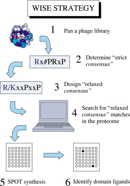
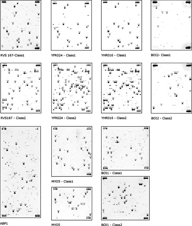
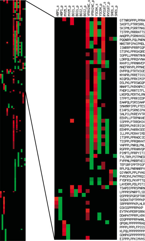
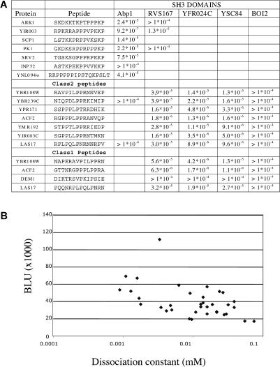
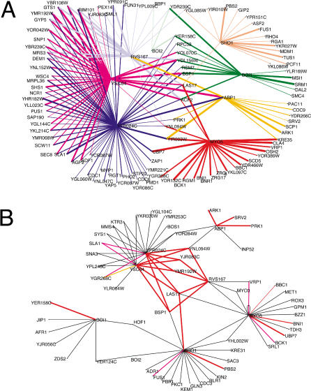
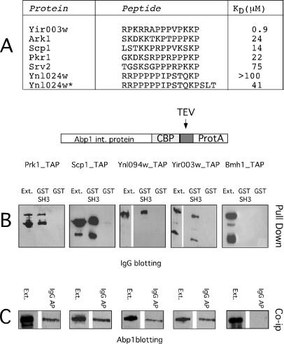
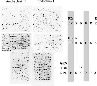
A substantial proportion of protein interactions relies on small domains binding to short peptides in the partner proteins. Many of these interactions are relatively low affinity and transient, and they impact on signal transduction. However, neither the number of potential interactions mediated by each domain nor the degree of promiscuity at a whole proteome level has been investigated. We have used a combination of phage display and SPOT synthesis to discover all the peptides in the yeast proteome that have the potential to bind to eight SH3 domains. We first identified the peptides that match a relaxed consensus, as deduced from peptides selected by phage display experiments. Next, we synthesized all the matching peptides at high density on a cellulose membrane, and we probed them directly with the SH3 domains. The domains that we have studied were grouped by this approach into five classes with partially overlapping specificity. Within the classes, however, the domains display a high promiscuity and bind to a large number of common targets with comparable affinity. We estimate that the yeast proteome contains as few as six peptides that bind to the Abp1 SH3 domain with a dissociation constant lower than 100 microM, while it contains as many as 50-80 peptides with corresponding affinity for the SH3 domain of Yfr024c. All the targets of the Abp1 SH3 domain, identified by this approach, bind to the native protein in vivo, as shown by coimmunoprecipitation experiments. Finally, we demonstrate that this strategy can be extended to the analysis of the entire human proteome. We have developed an approach, named WISE (whole interactome scanning experiment), that permits rapid and reliable identification of the partners of any peptide recognition module by peptide scanning of a proteome. Since the SPOT synthesis approach is semiquantitative and provides an approximation of the dissociation constants of the several thousands of interactions that are simultaneously analyzed in an array format, the likelihood of each interaction occurring in any given physiological settings can be evaluated. WISE can be easily extended to a variety of protein interaction domains, including those binding to modified peptides, thereby offering a powerful proteomic tool to help completing a full description of the cell interactome.


Conflict of interest statement

The authors have declared that no conflicts of interest exist.

Figures

Figure 1
Figure 1. Schematic Representation of the WISE Strategy
Figure 2
Figure 2. WISE Screening of the Binding Potential of Yeast SH3 Domains
Seven GST–SH3 domain fusion proteins were challenged with peptides that match different relaxed consensi: class 1 (R/K)xxPxxP and class 2 PxxPx(R/K) . The Myo5 SH3 domain was also tested with peptides matching (F/P/L/W/A/E)xx(W/Y/L/M/F/H)xxPxxP, while the Abp1 membrane contains peptides matching either xxPx(K/R)P or Pxxx(K/R)P. In the design of these relaxed patterns, we first aimed at defining regular expressions that could retrieve from the proteome all the peptides that had been demonstrated, to bind to the domain under consideration. Whenever the number of matching peptides did not exceed an arbitrary chosen threshold of 1,500, we used subjective considerations about sidechain similarities to further relax the search pattern. The three spots near the membrane corners contain peptides that bind to the anti-GST antibody. The intensity of these spots was used for normalization.
Figure 3
Figure 3. Comparison of the Phage Display Prediction and the Results of the SPOT Binding Test by the WISE Approach
The quantitative results of the experiments in Figure 1 are visualized with a graphical representation obtained with the tool EPCLUST available at http://ep.ebi.ac.uk/EP/EPCLUST. The PepSpot data, represented in red in a semiquantitative scale, is compared to the phage display prediction. Only peptides with BLUs (measured on a Lumi-ImagerTM) higher than 25K are included in the representation. The red intensity scale corresponds to BLU values in the ranges 25K–35K, 35K–45K, 45K–55K, 55K–85K, and larger than 85K, where higher BLU values correspond to a brighter red. Peptides that obtained a high score with the phage display-derived position-specific scoring matrix (Tong et al. 2002) are in brighter green. Peptides with a lower score are represented with a correspondingly lighter green according to an arbitrary linear scale.
Figure 4
Figure 4. Measurement of Dissociation Constants and Correlation with SPOT Intensities
(A) Dissociation constants were measured with a BIAcoreX instrument as described in the Materials and Methods. The experiments with the Abp1 SH3 domain were carried out in triplicate. (B) Normalized BLU intensities plotted as a function of the log of the dissociation constant.
Figure 5
Figure 5. Inferred Protein Interaction Networks
(A) Protein interaction network mediated by the SH3 domains of the proteins characterized in this study. The SH3-containing proteins are represented as blue dots, while the prey partner proteins are represented as black dots. The interactions mediated by each SH3 are represented in a different color, and the edge thicknesses are proportional to the BLU intensity of the corresponding interaction, according to the scale described in Figure 3. (B) The graph represents the interaction network mediated by the SH3 domains of Rvs167, Ysc84, Yfr024c, Abp1, Myo5, Sho1, Boi1, and Boi2 as determined by the two-hybrid approach (Tong et al. 2002). The interactions (edges) that were confirmed by our WISE method (BLU value higher than 25K) are colored in red or magenta. The interactions in magenta, differently from the ones in red, were not correctly inferred by the phage display approach. The interaction in orange was inferred by the phage display approach, but not confirmed by the WISE method. The network was visualized by the Pajek package (http://vlado.fmf.uni-lj.si/pub/networks/pajek/).
Figure 6
Figure 6. Characterization of Abp1 Ligands
(A) The dissociation constants of the 11 peptides that bound most efficiently to the Abp1 SH3 domain in the SPOT synthesis assay were measured by BIAcore experiments. (See also Table S1.) The results of the experiments for the peptides with the highest affinity are reported here. (B) The genes encoding the putative Abp1 ligands (Prk1, Yir003w, Scp1, and Ynl094w) were modified by the TAP technology to produce tagged proteins. A strain expressing the “tapped” Bmh1 protein is used as a control. Yeast extracts encoding the tagged proteins were used in pulldown experiments in the presence of 100 μg of GST–Abp1 SH3 or GST alone as a negative control. The “Ext.” lane was loaded with 1/20 of the extract used in the pulldown experiment. (C) The same extracts were affinity-purified on an IgG affinity resin and then the affinity tag, protein A, released by cutting with the TEV protease. The proteins that were copurified with the “tapped” baits were revealed with an anti-Abp1 serum.
Figure 7
Figure 7. Scanning of the Human Proteome in Search of Ligands for the Amphiphysin and Endophilin SH3 Domains
The relaxed target peptide consensi (right) were derived from the available phage display experimental data and used to search the human proteins contained in the SwissProt/TREMBL database with the software ScanProsite, found at http://us.expasy.org/tools/scanprosite/.

References

    1. Aasland R, Abrams C, Ampe C, Ball LJ, Bedford MT, et al. Normalization of nomenclature for peptide motifs as ligands of modular protein domains. FEBS Lett. 2002;513:141–144. - PubMed
    1. Barnett P, Bottger G, Klein AT, Tabak HF, Distel B. The peroxisomal membrane protein Pex13p shows a novel mode of SH3 interaction. EMBO J. 2000;19:6382–6391. - PMC - PubMed
    1. Cestra G, Castagnoli L, Dente L, Minenkova O, Petrelli A, et al. The SH3 domains of endophilin and amphiphysin bind to the proline-rich region of synaptojanin 1 at distinct sites that display an unconventional binding specificity. J Biol Chem. 1999;274:32001–32007. - PubMed
    1. de Heuvel E, Bell AW, Ramjaun AR, Wong K, Sossin WS, et al. Identification of the major synaptojanin-binding proteins in brain. J Biol Chem. 1997;272:8710–8716. - PubMed
    1. Fazi B, Cope MJ, Douangamath A, Ferracuti S, Schirwitz K, et al. Unusual binding properties of the SH3 domain of the yeast actin-binding protein Abp1: Structural and functional analysis. J Biol Chem. 2002;277:5290–5298. - PubMed

Publication types