. 2004 Jan 22:5:6.
doi: 10.1186/1471-2121-5-6.

Fas induces apoptosis in human coronary artery endothelial cells in vitro

Affiliations

Fas induces apoptosis in human coronary artery endothelial cells in vitro

Gerasimos Filippatos et al. BMC Cell Biol. .

Abstract

Background: Published work suggests that some types of endothelial cells undergo apoptosis in response to ligation of the receptor Fas (CD95, APO1) but other types are resistant. Because heterogeneity among endothelial cells from different tissues, has been demonstrated, the purpose of this study was to determine, if Fas ligation and/or activation by human Fas ligand induces apoptosis and caspase activities, in cultured human coronary artery endothelial cells, and the differences between TNF-a and FAS induced apoptosis in these cells.

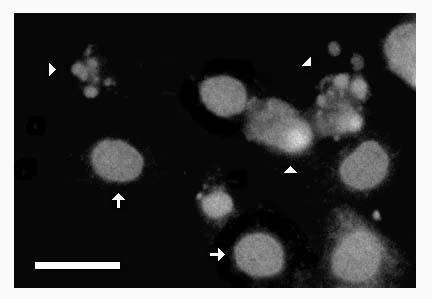
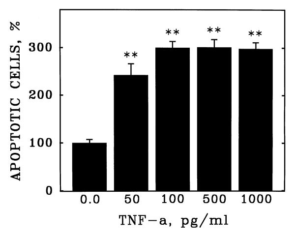
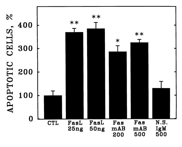
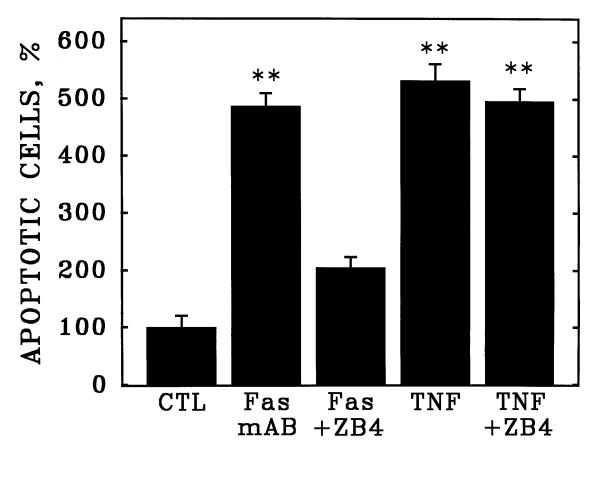
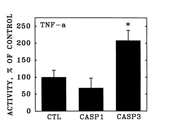
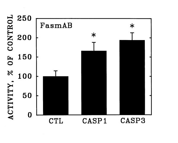
Results: Cultured human coronary artery endothelial cells (HCAEC) were exposed to the monoclonal Fas-activating antibody CH-11, to purified recombinant human Fas ligand, to the Fas-neutralizing antibody ZB4, or to purified recombinant human TNF-alpha. Apoptosis was detected by assessment of chromatin condensation and nuclear fragmentation and by assay of the enzymatic activities of Caspase 1 and Caspase 3 with membrane-permeable substrates applied to intact cells. Fas protein was detected by immunoblotting of HCAEC lysates. Apoptosis was induced in HCAEC by purified Fas ligand or by the monoclonal activating antibody CH-11 at concentrations of 25 or 200 ng/ml, but not by nonspecific isotype-matched immunoglobulins. The apoptotic index elicited by either Fas activator was equal to that induced by TNF-a (3.0-3.6-fold versus control, p < 0.01). The Fas-neutralizing antibody ZB4 abrogated HCAEC apoptosis induced by CH-11, but had no inhibitory effect on apoptosis in response to TNF-a. Fas ligation significantly increased the activities of both Caspase 1 and Caspase 3 at 20 hours of stimulation (1.7- and 2.0-fold versus control, both p < 0.05); in contrast, purified TNF-a increased the activity of Caspase 3 but not Caspase 1 (2.1-fold, p < 0.05). Western blotting of HCAEC lysates with antibody CH-11 identified a single immunoreactive protein of 90 kDa.

Conclusions: Cultured human coronary artery endothelial cells express functional Fas capable of inducing apoptosis in response to either purified Fas ligand or receptor-activating monoclonal antibodies, at levels equal to those inducible by purified TNF-alpha. Immunologic studies and differential kinetics of caspase activation suggest that Fas and TNF-alpha induce apoptosis in HCAEC by signaling pathways that are distinct but equal in potency.


Figures

Figure 1
Figure 1
Fluorescence detection of apoptosis in cultured human coronary artery endothelial cells. Human coronary artery endothelial cells (HCAEC) were incubated with purified TNF-α (1 ng/ml) in serum-free medium. After 20 hours, the culture vessel was centrifuged to retain detached cells and was labeled with propidium iodide (see Materials and Methods). Greyscale image depicts red fluorescence (>590 nm) due to chromatin-bound propidium iodide in ethanol-fixed HCAEC treated with DNAse-free RNAse. Note condensed chromatin in discrete nuclear fragments in apoptotic cells (arrowheads). Arrows depict normal nuclei. Bar = 10 microns.
Figure 2
Figure 2
Induction of apoptosis in cultured human coronary artery endothelial cells by purified human TNF-α. Human coronary artery endothelial cells (HCAEC) were incubated with purified recombinant human TNF-α at the indicated concentrations (ng/ml) in serum-free medium. After 20 hours, apoptosis was detected as described in Figure 1. Bars are the mean + S.E.M. of at least three determinations; ** = p < 0.01 versus 0.0 dose by ANOVA and Student-Newman-Keul's test.
Figure 3
Figure 3
Induction of apoptosis in cultured human coronary artery endothelial cells by activation of Fas. Human coronary artery endothelial cells (HCAEC) were incubated with purified recombinant human Fas ligand (FasL), Fas-activating monoclonal antibody CH-11 (Fas mAB), or nonspecific isotype-matched immunoglobulins (N.S.IgM) at the indicated concentrations (ng/ml) in serum-free medium. After 20 hours, apoptosis was detected as described in Figure 1. Bars are the mean + S.E.M. of at least three determinations; ** = p < 0.01 versus control (CTL) and * = p < 0.05 versus CTL by ANOVA and Student-Newman-Keul's test.
Figure 4
Figure 4
Identification of Fas antigen in human coronary artery endothelial cells. Cell lysates were prepared from cultured HCAEC and were analyzed by western blotting with the same anti-Fas monoclonal antibody used for receptor activation in Figure 3. Note single major immunoreactive band (arrowhead). STD = molecular mass standards in kDa.
Figure 5
Figure 5
Fas-neutralizing antibodies abrogate apoptosis of cultured human coronary artery endothelial cells in response to Fas but not TNF-α. Cultured HCAEC were incubated with Fas-activating monoclonal antibody CH-11 (Fas) or purified human TNF-α (TNF) in the presence or absence of the Fas-neutralizing monoclonal antibody, clone ZB4. Bars are the mean + S.E.M. of at least three determinations; ** = p < 0.01 versus control (CTL) by ANOVA and Student-Newman-Keul's test.
Figure 6
Figure 6
Stimulation of Caspase 3 activity in human coronary artery endothelial cells by purified TNF-α. Cultured human coronary artery endothelial cells were incubated for 20 hours in the presence and absence of purified recombinant TNF-α and were then detached from the culture surfaces. Over the subsequent hour, the cells were incubated in suspension culture with membrane-permeable fluorescent substrates specific for Caspase 1 (CASP1) and Caspase 3 (CASP3). Data are expressed as substrate cleaved per 106 cells, relative to unstimulated control cultures (CTL). Bars are the mean + S.E.M. of at least three determinations; * = p < 0.05 versus unstimulated control (CTL) by ANOVA and Student-Newman-Keul's test.
Figure 7
Figure 7
Stimulation of Caspase 1 and Caspase 3 activities in human coronary artery endothelial cells by activation of Fas. Cultured human coronary artery endothelial cells were incubated for 20 hours in the presence and absence of anti-Fas clone CH-11 activating antibody, and were then detached from the culture surfaces. Over the subsequent hour, the activities of Caspase 1 (CASP1) and Caspase 3 (CASP3) were determined as described in Figure 6. Data are expressed as substrate cleaved per 106 cells, relative to unstimulated control cultures (CTL). Bars are the mean + S.E.M. of at least three determinations; * = p < 0.05 versus unstimulated control (CTL) by ANOVA and Student-Newman-Keul's test.

Similar articles

Cited by

References

    1. Abrams J. Role of endothelial dysfunction in coronary artery disease. Am J Cardiology. 1997;79:2–9. doi: 10.1016/S0002-9149(97)00379-2. - DOI - PubMed
    1. Rubanyi GM. The role of endothelium in cardiovascular homeostasis and diseases. J Cardiovasc Pharmacol. 1993;22:S1–S14. - PubMed
    1. Ruschitza F, Noll G, Luscher T. The endothelium in coronary artery disease. Cardiology. 1997;88:3–19. - PubMed
    1. Geng YJ, Libby P. Evidence for apoptosis in advanced human atheroma: colocalization with interleukin-1b-converting enzyme. Am J Pathol. 1995;147:251–266. - PMC - PubMed
    1. Bochaton-Piallat M, Gabbiani F, Redard M, Desmouliere A, Gabbiani G. Apoptosis participates in cellular regulation during rat aortic intimal thickening. Am J Pathol. 1995;146:1059–1064. - PMC - PubMed

Publication types