. 2004 Jan 22;32(2):e19.
doi: 10.1093/nar/gnh014.

Construction of long DNA molecules using long PCR-based fusion of several fragments simultaneously

Affiliations

Construction of long DNA molecules using long PCR-based fusion of several fragments simultaneously

Nikolai A Shevchuk et al. Nucleic Acids Res. .

Abstract

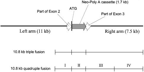
A procedure for precise assembly of linear DNA constructs as long as 20 kb is proposed. The method, which we call long multiple fusion, has been used to assemble up to four fragments simultaneously (for a 10.8 kb final product), offering an additional improvement on the combination of long PCR and overlap extension PCR. The method is based on Pfu polymerase mix, which has a proofreading activity. We successfully assembled (and confirmed by sequencing) seven different linear constructs ranging from 3 to 20 kb, including two 20 kb products (from fragments of 11, 1.7 and 7.5 kb), two 10.8 kb constructs, and two constructs of 6.1 and 6.2 kb, respectively. Accuracy of the PCR fusion is greater than or equal to one error per 6.6 kb, which is consistent with the expected error rate of the PCR mix. The method is expected to facilitate various kinds of complex genetic engineering projects that require precise in-frame assembly of multiple fragments, such as somatic cell knockout in human cells or creation of whole genomes of viruses for vaccine research.


Figures

Figure 1
Figure 1
Outline of long triple fusion (sizes of fragments are not to scale).
Figure 2
Figure 2
Mechanism of triple fusion at step A (primers are added at step B). Individual strands of DNA fragments and their 5′ and 3′ ends are shown. The flow chart shows theoretical stages of formation of double and triple fusion products. Each stage roughly corresponds to a PCR cycle (denaturation, annealing and extension of primed 3′ ends). Overlapping ends (of identical sequence) of fragments are shown with identical shading. Note that complementary ends have different fill patterns in the schematic. Triple fusion products are then amplified using nested primers at step B (not shown).
Figure 3
Figure 3
Outline of long quadruple fusion (not to scale).
Figure 4
Figure 4
Analysis of the product of triple fusion of three 1 kb fragments in 1% agarose gel. Lane 1, PCR product; lane 2, DNA molecular weight markers. Note that primers used in step B were not nested.
Figure 5
Figure 5
Electrophoretic analysis of the starting material and a 10.8 kb product of long triple fusion in 0.5% agarose gel. Lane 1, 1.9 kb left arm; lane 2, 1.7 kb Neo-polyA cassette (second fragment); lane 3, DNA 1 kb step ladder (Promega), 2–10 kb; lane 4, 7.5 kb right arm; lane 5, 10.8 kb triple fusion product. Length of the product is not the same as the sum of initial fragments because of the presence of overlaps and use of nested primers.
Figure 6
Figure 6
Various long triple fusion products. (A) Pulse-field electrophoresis. Lane 1, 20 kb product of fusion of fragments 11.2, 1.7 (Neo-polyA) and 7.5 kb; lane 2, 10.8 kb product of fusion of fragments 1.8, 1.7 (Neo-polyA) and 7.5 kb; lane 3, high molecular weight markers 8.3–48.5 kb (Bio-Rad), from bottom to top: 12.2, 15.0, 17.1, 19.4, 22.6, 24.8, 29.9, 33.5, 38.4 and 48.5; lane 4, 20 kb product of fusion of fragments 11.2, 1.7 (Hygro-polyA) and 7.5 kb. (B) Electrophoresis in 1% agarose (Sigma). Lane 1, 1 kb DNA step ladder (Promega), 1–10 kb; lane 2, 20 kb Neo-polyA product.
Figure 7
Figure 7
The structure of the neomycin-based 20 kb recombinant product. The locations of the corresponding fragments used for 10.8 kb triple fusion (Fig. 6A, lane 2) and 10.8 quadruple fusion (see Figs 3 and 9) are shown.
Figure 8
Figure 8
The structure of the hygromycin-based 20 kb recombinant product (see also Fig. 6A, lane 4).
Figure 9
Figure 9
Electrophoretic analysis of the starting material and a 10.8 kb product of long quadruple fusion in 0.5% agarose gel. Lane 1, 1.9 kb first fragment; lane 2, 1.7 kb Neo-polyA cassette (second fragment); lane 3, 3.6 kb third fragment; lane 4, 3.9 kb fourth fragment; lane 5, DNA 1 kb step ladder (Promega), 1–10 kb; lane 6, 10.8 kb recombinant product. Length of the product is not the same as the sum of initial fragments, because of overlaps and use of nested primers.
Figure 10
Figure 10
Structure of vectors GFP One (upper panel) and GFP Two (lower panel). Each of these constructs was synthesized from three fragments: the left arm (up to exon 3 start, ∼2.3 kb), startless GFP (680 bp) and the right arm (∼3.6 kb). Exons and introns are from human GM3 synthase gene. See also Figure 11.
Figure 11
Figure 11
Assembly of GFP-based targeting vectors (vectors GFP One and GFP Two in Fig. 10). Left: amplification of components from non-isogenic genomic DNA (homologous regions) and phMGFP plasmid vector (GFP protein coding region). Lane 1, GFP fragment (680 bp) amplified with chimeric primers (85 nt each) specific for targeting vector One; lane 2, GFP fragment (680 bp) amplified with chimeric primers (85 nt each) specific for targeting vector Two; lane 3, left homologous arm (2.3 kb, the same for both vectors); lane 4, right homologous arm (3.6 kb) for vector One; lane 5, right homologous arm (3.6 kb) for vector Two; lane M, DNA molecular weight markers, 1–10 kb. Right: PCR products obtained using long triple fusion method. Lane 1, targeting vector GFP One, 6.1 kb (see Fig. 10, upper panel); lane 2, targeting vector GFP Two, 6.2 kb (see Fig. 10, lower panel); lane M, DNA molecular weight markers, 1–10 kb.
Figure 12
Figure 12
Sample sequence of a fusion point in 6.2 kb construct GFP Two. Expected sequence is shown as letters beneath peaks. Color code: green, A; blue, C; black, G; red, T. Double underscored region, end of intron 2; asterisk, start of exon 3; single underscored region, beginning of startless Monster GFP open reading frame. See also Figure 10, lower panel and Supplementary Material, folder 6, file NS2-SQGFP1.ab1, starting from position 48.

References

    1. Yon J. and Fried,M. (1989) Precise gene fusion by PCR. Nucleic Acids Res., 17, 4895. - PMC - PubMed
    1. Yolov A.A. and Shabarova,Z.A. (1990) Constructing DNA by polymerase recombination. Nucleic Acids Res., 18, 3983–3986. - PMC - PubMed
    1. Pont-Kingdon G. (1997) Creation of chimeric junctions, deletions, and insertions by PCR. Methods Mol. Biol., 67, 167–172. - PubMed
    1. Kuwayama H., Obara,S., Morio,T., Katoh,M., Urushihara,H. and Tanaka,Y. (2002) PCR-mediated generation of a gene disruption construct without the use of DNA ligase and plasmid vectors. Nucleic Acids Res., 30, e2. - PMC - PubMed
    1. Sedivy J.M., Vogelstein,B., Liber,H.L., Hendrickson,E.A. and Rosmarin,A. (1999) Gene targeting in human cells without isogenic DNA. Science, 283, 9a.

Publication types