. 2004 Feb 3;101(5):1257-62.
doi: 10.1073/pnas.0308090100. Epub 2004 Jan 22.

Cooperation of the ErbB2 receptor and transforming growth factor beta in induction of migration and invasion in mammary epithelial cells

Affiliations

Cooperation of the ErbB2 receptor and transforming growth factor beta in induction of migration and invasion in mammary epithelial cells

Sarah E Seton-Rogers et al. Proc Natl Acad Sci U S A. .

Abstract

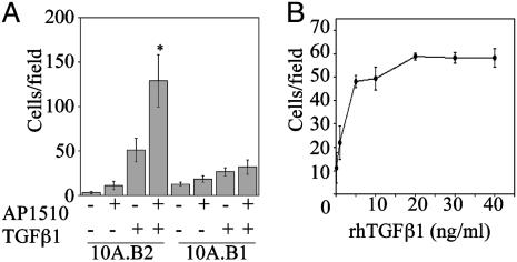
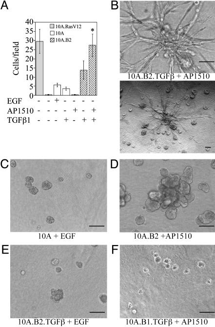
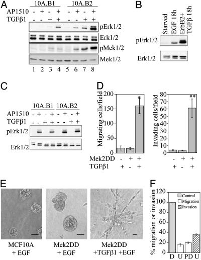
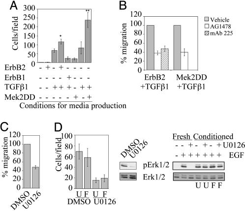
MCF10A mammary epithelial cells form growth-arrested structures when cultured in three-dimensional basement membrane gels. Activation of the receptor tyrosine kinase ErbB2 induces formation of proliferative structures that share properties with noninvasive early stage lesions. We conducted a genetic screen to identify cDNAs that can cooperate with ErbB2 to induce migration in these cells, with the hypothesis that they would represent candidate "second hits" in the development of invasive breast carcinomas. We found that expression of transforming growth factor (TGF)beta1 and TGFbeta3 in cells expressing activated ErbB2 induces migration in transwell chambers and invasive behavior in both basement membrane cultures and invasion chambers. The ability of ErbB2 to cooperate with TGFbeta correlated with sustained, elevated activation of extracellular signal-regulated kinase (Erk)-mitogen-activated protein kinase. Pharmacological reduction of Erk activity inhibited the cooperative effect of TGFbeta and ErbB2 on migration and expression of activated Erk kinase was sufficient to cooperate with TGFbeta to induce migration and invasion, suggesting that sustained Erk activation is critical for ErbB2/TGFbeta cooperation. In addition, we show that costimulation of ErbB2 and TGFbeta induces autocrine secretion of factors that are sufficient to induce migration, but not invasion, by means of both epidermal growth factor receptor-dependent and -independent processes. These results support the role of TGFbeta as a pro-invasion factor in the progression of breast cancers with activated ErbB2 and suggest that activation of the Erk and epidermal growth factor receptor pathways are key in mediating these events.


Figures

Fig. 1.
Fig. 1.
ErbB2 and TGFβ cooperate to induce migration of MCF10A cells. (A) Cells (10A.B2 or 10A.B1) expressing TGFβ1 or control vector were seeded in transwell migration chambers –/+ AP1510, incubated for 18 h, and quantified as detailed in Materials and Methods. *, P < 0.0001 and P = 0.048 as compared with ErbB2 or TGFβ alone, respectively. (B) Cells (10A.B2) were seeded in transwell chambers in the presence of 500 nM AP1510 and indicated doses of rhTGFβ1 for 18 h and quantified as for A.
Fig. 2.
Fig. 2.
ErbB2 and TGFβ cooperate to induce invasion. (A) Cells (10A.B2 and 10A.B2.TGFβ; hatched bars) were seeded in transwell invasion chambers with or without AP1510 and incubated for 24 h. MCF10A cells expressing RasV12 (10A.RasV12; gray bar) served as a positive control and MCF10A cells with or without EGF or TGFβ (white bars) as negative controls. *, P = 0.0023 and 0.0188 as compared with ErbB2 or TGFβ (in 10A.B2 cells) alone, respectively. (B–F) Representative three-dimensional structures of 10A.B2.TGFβ (B) cells treated with 500 nM AP1510; the same structure is shown at high (Upper) and low (Lower) magnification. MCF10A cells plus EGF (C), 10A.B2 cells plus AP1510 (D), 10A.B2.TGFβ cells plus EGF (E), and 10A.B1.TGFβ cells plus AP1510 (F). Structures were photographed at day 17. (Bars, 100 μm.)
Fig. 3.
Fig. 3.
Role of the Erk pathway in migration and invasion induced by ErbB2 and TGFβ. (A) Immunoblot analysis of phosphorylated Erk1/2 (pErk1/2) and Mek1/2 (pMek1/2) in 10A.B1 and 10A.B2 cells with or without TGFβ expression and AP1510 treatment for 18 h. (B) pErk1/2 levels in 10A.B2 cells treated with 20 ng/ml EGF or with TGFβ expression and AP1510 treatment for 18 h. (C) pErk1/2 levels in 10A.B1 and 10A.B2 cells with or without TGFβ expression and AP1510 treatment for 15 min. (D) MCF10A cells expressing Mek2DD or control vector were analyzed in transwell migration (Left) and invasion (Right) assays with or without TGFβ expression. *, P < 0.0002; **, P < 0.001 as compared with either Mek2DD or TGFβ alone. (E) Representative three-dimensional structures of MCF10A cells expressing control vector, Mek2DD, or Mek2DD plus TGFβ from day 15. (Bars, 40 μm.) (F) Transwell migration and invasion assays were performed with 10A.B2.TGFβ cells in the presence of AP1510 and Mek inhibitors U0126 (U; 5 μM) or PD98059 (PD; 50 μM), or DMSO control (D). Data are expressed as the percent of control cells (gray bar, normalized to 100%) that migrate (white bars) or invade (hatched bar) in the presence of inhibitor. PD98059 was not tested in invasion.
Fig. 4.
Fig. 4.
The combined activation of ErbB2 plus TGFβ or Mek2 plus TGFβ leads to secretion of both EGFR-dependent and -independent soluble migratory factors. (A) MCF10A cells were seeded in transwell migration chambers, and conditioned media produced from cells as indicated was added to the bottom chambers and incubated for 18 h. *, P = 0.0056 as compared with TGFβ alone; P = 0.004 as compared with ErbB1 plus TGFβ. **, P = 0.008 as compared with TGFβ alone; P = 0.002 as compared with MEK2DD alone. (B) Migration assays were performed as described in A, except EGFR inhibitors AG1478 (300 nM) or mAb 225 (10 μg/ml) were added. Data are expressed as the percent of control cells that migrate or invade with inhibitor. Migration with Mek 2DD plus TGFβ-conditioned medium was not tested with mAb 225. (C) Migration assays were performed by using 10A.B2.TGFβ plus AP1510-conditioned medium and 5 μM U0126. Data are expressed as the percent of control cells that migrate with inhibitor. (D) Conditioned media from 10A.B2.TGFβ cells treated with AP1510 was made in the presence of 5 μM U0126 or DMSO and inhibitor removed by filtration. Transwell migration assays were performed with this media and MCF10A cells (Left; U, unfiltered medium; F, filtered medium). To show inhibition of Erk phosphorylation by U0126 in the cells used to make the media, they were lysed after media collection and pErk1/2 levels were analyzed by immunoblot (Center). To show that migration inhibition was not due to carryover of U0126 after filtration, MCF10A cells were pretreated for 15 min with conditioned medium or fresh U0126 and were stimulated for 15 min with 20 ng/ml EGF. Lysates of these cells were analyzed by immunoblotting pErk1/2 (Right; U, unfiltered medium; F, filtered medium).

References

    1. Harari, D. & Yarden, Y. (2000) Oncogene 19, 6102–6114. - PubMed
    1. Hynes, N. E. & Stern, D. F. (1994) Biochim. Biophys. Acta 1198, 165–184. - PubMed
    1. Olayioye, M. A., Neve, R. M., Lane, H. A. & Hynes, N. E. (2000) EMBO J. 19, 3159–3167. - PMC - PubMed
    1. Di Fiore, P. P., Pierce, J. H., Kraus, M. H., Segatto, O., King, C. R. & Aaronson, S. A. (1987) Science 237, 178–182. - PubMed
    1. Samanta, A., LeVea, C. M., Dougall, W. C., Qian, X. & Greene, M. I. (1994) Proc. Natl. Acad. Sci. USA 91, 1711–1715. - PMC - PubMed

Publication types

Substances