. 2004 Feb 3;101(5):1333-8.
doi: 10.1073/pnas.0308125100. Epub 2004 Jan 22.

Differentiation and developmental pathways of uropathogenic Escherichia coli in urinary tract pathogenesis

Affiliations

Differentiation and developmental pathways of uropathogenic Escherichia coli in urinary tract pathogenesis

Sheryl S Justice et al. Proc Natl Acad Sci U S A. .

Abstract

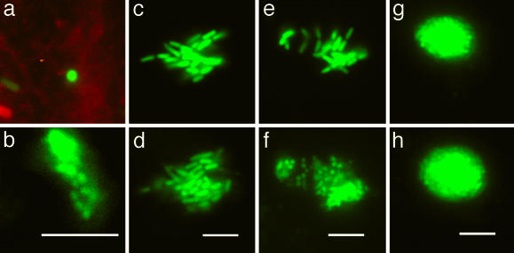
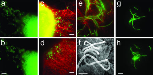
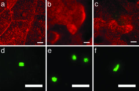
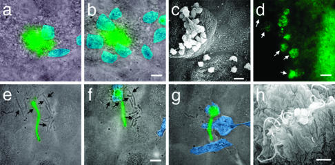
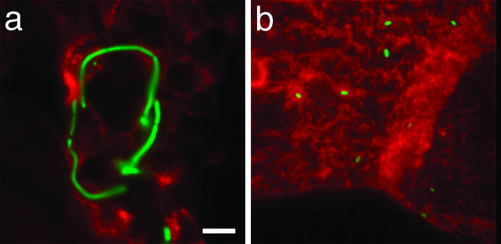
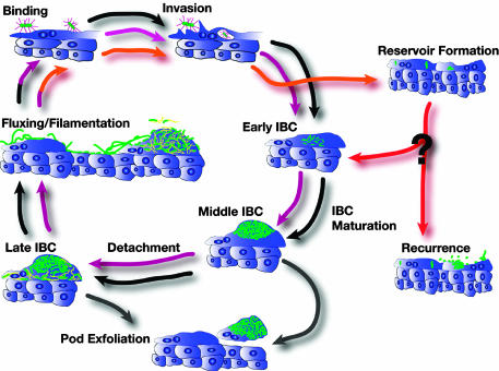
Uropathogenic Escherichia coli (UPEC) are capable of forming complex intracellular bacterial communities (IBC) within the superficial umbrella cells of the bladders of C3H and BALB/c mice. By using time-lapse fluorescence videomicroscopy to observe infected mouse bladder explants, we discovered that IBCs formed by uropathogenic E. coli progressed through four distinct developmental stages that differed with respect to growth rate, bacterial length, colony organization, motility, and its eventual dispersal. In the first phase, bacteria in the IBC were nonmotile, rod shaped, and grew rapidly in loosely organized colonies free in the cytoplasm of the bladder superficial umbrella cells. In the second phase, the loose collection of bacteria in the IBC matured into a slower growing, highly organized biofilm-like community consisting of coccoid bacteria that ultimately filled most of the cytoplasm. In the third phase, bacteria in the biofilm-like state in the IBC switched to a motile rod-shaped phenotype allowing detachment from the community and eventual fluxing out of the host cell. During the fourth phase, the bacteria filamented. Filamentation appeared to be in response to a Toll-like receptor 4-mediated innate defense mechanism. Bacteria that fluxed out of the superficial umbrella cells were able to reenter the IBC developmental cascade but with slower kinetics and ultimately a quiescent reservoir was established. Intracellular growth and filamentation provided an advantage to the bacteria in evading infiltrating polymorphonuclear leukocytes. This work has developed a technique to observe live infected organs and revealed a complex differentiation pathway that facilitates bacterial persistence in the urinary tract.


Figures

Fig. 1.
Fig. 1.
Maturation of an IBC. These micrographs depict three stages of the developmental program of UPEC during the acute infection. GFP-producing bacteria were visualized by fluorescence videomicroscopy to monitor the events in real time by using r-WGA as a cell surface marker as described in the text. Each pair of micrographs depicts events observed in different superficial umbrella cells. Animated versions are available in Movies 1–3, which are published as supporting information on the PNAS web site. (a) At 2 h after infection, a single bacterium (green) is perpendicular to the bladder cell surface (red) and invades the cell over 10 min. (b) After 12 h, this bacterium formed an intracellular microcolony ≈2 μm below the surface. (c and d) The morphology and growth of the early IBC are depicted as snapshots taken 42 min apart. (e and f) Maturation to the middle IBC is depicted in snapshots taken 100 min apart. (g and h) Growth and architecture of the middle IBC are depicted as snapshots taken 42 min apart. (Scale bars = 10 μm.)
Fig. 2.
Fig. 2.
Fluxing and filamentation. (a and b) Detachment and fluxing are depicted as snapshots taken 3 s apart. Detached bacteria are swimming rapidly. Animated versions are available in Movies 4 and 5, which are published as supporting information on the PNAS web site. (c and d) Two representative fluorescence micrographs depicting escaped bacteria (green) on the surface of two different superficial umbrella cells (red). The bright yellow patch (c) is the epifluorescence of the IBC within the superficial umbrella cell. Abundance of filamentous bacteria on the bladder surface is depicted by fluorescence micrograph (e) and by SEM (f). (g and h) The release of daughter cells from the filaments and their subsequent growth are depicted as snapshots taken 36 min apart. (Scale bars = 10 μm in bd and h and 5 μm in f.)
Fig. 3.
Fig. 3.
Massive exfoliation and quiescent reservoir. (ac) r-WGA staining of infected mouse bladder showed the presence of small, regenerating bladder epithelial cells after a massive exfoliation. (df) Fluorescence micrographs depicting bacteria within the superficial umbrella cells after exfoliation. The majority of the bacteria were pairs (d), small clusters (e), or a pair where one bacterium was longer than the other (f). (Scale bars = 10 μm.)
Fig. 4.
Fig. 4.
Pod protects UPEC from PMN attack. Fluorescence micrographs were overlaid with a transmitted light image taken at the same time. The PMNs were pseudocolored blue for ease of visualization. Animated versions are available in Movies 6 –9, which are published as supporting information on the PNAS web site. (a) From the surface, the epifluorescence of the IBC was observed (green) while PMNs (blue) swarm on the surface. (b) Ten minutes later, more PMNs have been recruited but fail to gain access to the IBC. (c) SEM of bladder surface showing the presence of numerous PMNs on the surface of infected cells but absent from uninfected cells. (d) Fluorescence micrograph depicting PMNs consuming the IBC (white arrows). (eg) Overlay of the transmitted light image on the fluorescence micrograph depicting snapshots during which PMNs consume the dividing nonfluorescent bacteria (black arrows) but fail to consume the fluorescent filament. These frames were taken 13 min apart. (h) SEM of the bladder surface showing that filaments survive in the presence of PMNs. (Scale bars = 10 μm.)
Fig. 5.
Fig. 5.
Filamentation requires functional TLR-4. Filaments are observed on the luminal surface of the bladder from wild-type mice (C3H/HeN) (a) but are absent at the same time point in TLR-4-deficient mice (C3H/HeJ) (b).
Fig. 6.
Fig. 6.
UTI pathogenic cascade model. This model depicts the sequence of events during the progression of establishment of a UTI based on the data presented in this report in conjunction with previous studies. The first round of the developmental process (black arrows) directly leads to the second round (magenta arrows) that completes at the time of massive exfoliation of the superficial umbrella cells. At this time, the reservoir is established (orange arrows). Exfoliation of epithelial cells occurs as a mechanism of the innate immune system (gray arrows). Events that lead to recurrence and the mechanism of bacterial growth during recurrence are unclear (red arrows and question mark); these events may include reentry into the characterized cycle at the point of early IBC formation. Bacteria (green) bind to and invade into superficial umbrella cells via type 1 pili (purple). Detachment from IBC and fluxing out of infected cells likely involve flagella expression (yellow).

References

    1. Hooton, T. M. & Stamm, W. E. (1997) Infect. Dis. Clin. N. Am. 11, 551–581. - PubMed
    1. Svanborg, C. & Godaly, G. (1997) Infect. Dis. Clin. N. Am. 11, 513–529. - PubMed
    1. Foxman, B. (2002) Am. J. Med. 113, Suppl 1A, 5S–13S. - PubMed
    1. Foxman, B. (1990) Am. J. Public Health 80, 331–333. - PMC - PubMed
    1. Mysorekar, I. U., Mulvey, M. A., Hultgren, S. J. & Gordon, J. I. (2002) J. Biol. Chem. 277, 7412–7419. - PubMed