. 2004 Jan 28;23(2):294-301.
doi: 10.1038/sj.emboj.7600063. Epub 2004 Jan 22.

Escherichia coli YidC is a membrane insertase for Sec-independent proteins

Affiliations

Escherichia coli YidC is a membrane insertase for Sec-independent proteins

Justyna Serek et al. EMBO J. .

Abstract

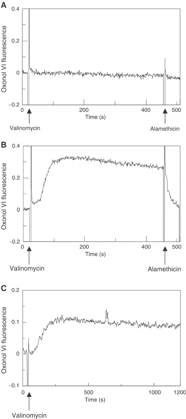
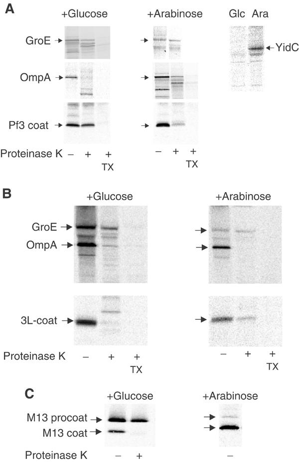
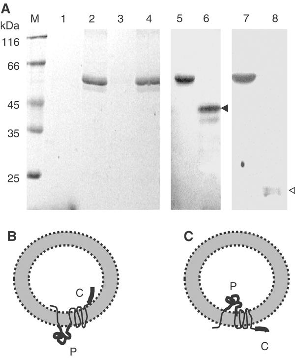
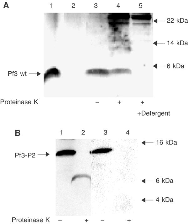
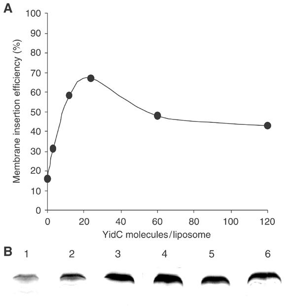
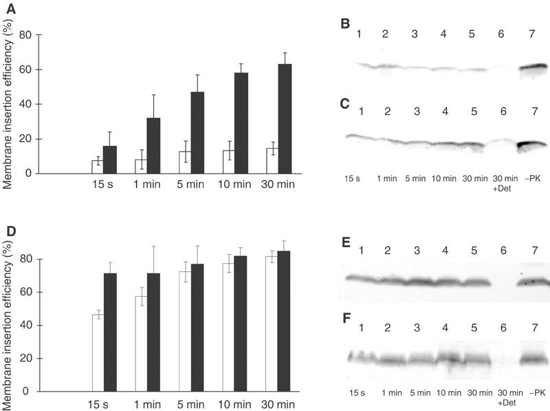
YidC is a recently discovered bacterial membrane protein that is related to the mitochondrial Oxa1p and the Alb3 protein of chloroplasts. These proteins are required in the membrane integration process of newly synthesized proteins that do not require the classical Sec machinery. Here we demonstrate that YidC is sufficient for the membrane integration of a Sec-independent protein. Microgram amounts of the purified single-spanning Pf3 coat protein were efficiently inserted into proteoliposomes containing the purified YidC. A mutant Pf3 coat protein with an extended hydrophobic region was inserted independently of YidC into the membrane both in vivo and in vitro, but its insertion was accelerated by YidC. These results show that YidC can function separately from the Sec translocase to integrate membrane proteins into the lipid bilayer.


Figures

Figure 1
Figure 1
Insertion of Pf3 coat protein into liposomes. Purified Pf3 coat protein (A, B) and purified 3L-Pf3 coat protein (C, D) were added to E. coli liposomes with a 0.25 μm mean diameter, generated with an extruder. The reactions were incubated at 37°C for 1 h and pelleted at 130 000 g. The Pf3 coat protein was found binding to the liposomes (lane 1). An aliquot was digested with 0.5 mg/ml proteinase K at 0°C for 30 min in the absence (lane 2) or presence of detergent (lane 3). The proteins were applied to liposomes (A, C) and energized liposomes (B, D). The samples were acid precipitated, analysed by PAGE and visualized by silver stain.
Figure 2
Figure 2
Generation of a membrane potential across liposomes. Extruded liposomes generated in 100 mM Na2SO4 were sedimented and resuspended in 100 mM Na2SO4 (A) or in 100 mM K2SO4 (B). To the liposomes, 0.1 μM oxonol VI was added and the fluorescence at an excitation/emission of 599/634 nm was recorded. After addition of 0.25 μM valinomycin, a transmembrane potential was generated as measured by the increase of the oxonol fluorescence. The membrane potential was abolished by the addition of 3 μg/ml alamethicin. The membrane potential was also monitored with YidC proteoliposomes (C).
Figure 3
Figure 3
Membrane insertion of the Pf3 coat protein in vivo. Protease mapping of the Pf3 coat protein (A) and the mutant 3L-Pf3 (B) expressed in JS 7131 cells depleted of YidC (+glucose, left panels) or expressing YidC (+arabinose, right panels). Exponentially growing cells bearing the respective plasmids were induced for 10 min with 1 mM IPTG and pulse labelled with [35S]-methionine for 3 min, converted to spheroplasts and digested with 200 μg/ml proteinase K either in the absence or presence of 2% Triton X-100. The samples were immunoprecipitated with antibodies to Pf3 phage (A, B, lower panels) or to OmpA, GroEL and YidC (upper panels) and analysed by SDS–PAGE and phosphorimaging. For a control, the processing and protease accessibility of the M13 procoat protein was analysed (C).
Figure 4
Figure 4
Reconstitution of YidC into proteoliposomes. (A) Purified YidC protein (lane 2) was mixed with E. coli lipids to form proteoliposomes. The proteoliposomes were pelleted in an airfuge (lane 4). The absence of the protein in the supernatant (lane 3) showed that it was efficiently integrated into the YidC-containing proteoliposomes. The samples were analysed by SDS–PAGE and Coomassie stained. For reference, molecular weight marker (lane M) and E. coli lipid (lane 1) were applied on the gel. The samples were analysed by SDS–PAGE and immunoblotted with an antibody directed to the periplasmic region of YidC (lanes 5 and 6) or to the C-terminal tail of YidC (lanes 7 and 8). Trypsin treatment of the proteoliposomes generated a 42 kDa fragment (lane 6, black arrowhead) or a 20 kDa fragment (lane 8, white arrowhead), respectively. (B, C) Two possible topologies of the reconstituted YidC. The large periplasmic domain (P) and the C-terminal tail (C) are highlighted.
Figure 5
Figure 5
Membrane insertion of Pf3 and Pf3-P2 coat proteins into YidC proteoliposomes in vitro. (A) Purified Pf3 coat protein (lane 1) was mixed with YidC proteoliposomes (lanes 2–5), incubated for 1 h at 37°C (lanes 3–5) and sedimented by centrifugation. Essentially all the protein was bound to the proteoliposomes (lane 2 supernatant, lane 3 pellet). An aliquot of the sample was digested with 0.5 mg/ml proteinase K for 1 h in the absence (lane 4) or presence of detergent (lane 5). The samples were acid precipitated, and analysed by SDS–PAGE and silver staining. The insertion efficiency of the Pf3 coat protein was quantified as 74% on calculating the difference between the Pf3 coat protein bands in lanes 3 and 4 using a Kodak imager. (B) Purified Pf3-P2 was added to YidC proteoliposomes as in (A) (lanes 1 and 3), and analysed by proteinase K (lanes 2 and 4). The samples were applied to SDS–PAGE and immunoblotted with antiserum to the N-terminal region of the Pf3 protein (lanes 1 and 2) or to leader peptidase P2 region (lanes 3 and 4). For reference, the positions of a molecular weight marker are indicated by arrows.
Figure 6
Figure 6
Proteoliposomes with different YidC contents. Proteoliposomes were generated that had YidC:lipid ratios varying from 1:5000 to 1:200 000. Assuming 650 000 lipid molecules per liposome, this gives a range from 3 to 120 YidC molecules per liposome (A). Liposomes without YidC (B, lane 1) and proteoliposomes of YidC:lipid ratios of 1:200 000 (lane 2), 1:50 000 (lane 3), 1:25 000 (lane 4), 1:10 000 (lane 5) and 1:5000 (lane 6) were used to examine the membrane insertion of Pf3 coat following the identical protocol as in Figure 5. The efficiency of membrane insertion was quantified using a Kodak imager.
Figure 7
Figure 7
Kinetics of YidC-mediated membrane insertion. Reconstituted YidC proteoliposomes (A, D: black columns; panels C, F) or liposomes (A, D: white columns; panels B, E) were mixed with Pf3 coat protein (A–C) or the mutant 3L-Pf3 (D–F), and incubated at 37°C for the indicated times. The reaction was stopped by chilling on ice and the addition of 0.5 mg/ml proteinase K. The proteinase reaction was continued for 1 h at 0°C. The samples were then acid precipitated and analysed by a Western immunoblot with an antiserum to Pf3 phage. The bands were quantified as described in Figure 6. The mean values of five independent experiments were calculated (A, D).

References

    1. Akimaru J, Matsuyama S, Tokuda H, Mizushima S (1991) Reconstitution of a protein translocation system containing purified SecY, SecE, and SecA from Escherichia coli. Proc Natl Acad Sci USA 88: 654–659 - PMC - PubMed
    1. Apell HJ, Bersch B (1987) Oxonol VI as an optical indicator for membrane potentials in lipid vesicles. Biochim Biophys Acta 903: 480–494 - PubMed
    1. Bassilana M, Wickner W (1993) Purified Escherichia coli preprotein translocase catalyzes multiple cycles of precursor protein translocation. Biochemistry 32: 2626–2630 - PubMed
    1. Beck K, Wu LF, Brunner J, Müller M (2000) Discrimination between SRP- and SecA/SecB-dependent substrates involves selective recognition of nascent chains by SRP and trigger factor. EMBO J 19: 134–143 - PMC - PubMed
    1. Brundage L, Hendrick JP, Schiebel E, Driessen AJM, Wickner W (1990) The purified E. coli integral membrane protein SecY/E is sufficient for reconstitution of SecA-dependent precursor protein translocation. Cell 62: 649–657 - PubMed

Publication types

MeSH terms