. 2004 Feb;74(2):357-66.
doi: 10.1086/381685. Epub 2004 Jan 20.

Mutations in the VLGR1 gene implicate G-protein signaling in the pathogenesis of Usher syndrome type II

Affiliations

Mutations in the VLGR1 gene implicate G-protein signaling in the pathogenesis of Usher syndrome type II

Michael D Weston et al. Am J Hum Genet. 2004 Feb.

Erratum in

  • Am J Hum Genet. 2004 May;74(5):1080

Abstract

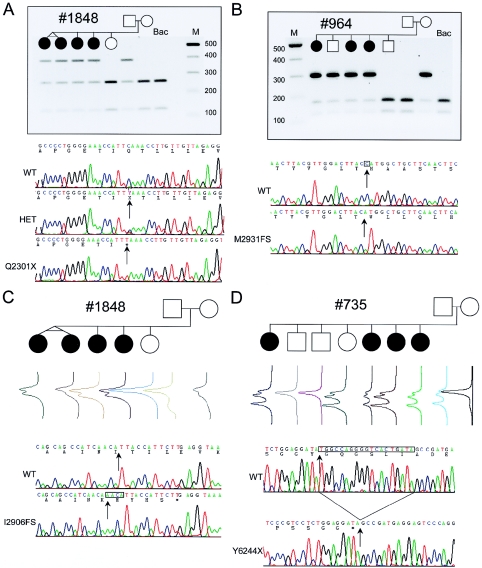
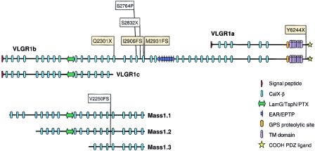
Usher syndrome type II (USH2) is a genetically heterogeneous autosomal recessive disorder with at least three genetic subtypes (USH2A, USH2B, and USH2C) and is classified phenotypically as congenital hearing loss and progressive retinitis pigmentosa. The VLGR1 (MASS1) gene in the 5q14.3-q21.1 USH2C locus was considered a likely candidate on the basis of its protein motif structure and expressed-sequence-tag representation from both cochlear and retinal subtracted libraries. Denaturing high-performance liquid chromatography and direct sequencing of polymerase-chain-reaction products amplified from 10 genetically independent patients with USH2C and 156 other patients with USH2 identified four isoform-specific VLGR1 mutations (Q2301X, I2906FS, M2931FS, and T6244X) from three families with USH2C, as well as two sporadic cases. All patients with VLGR1 mutations are female, a significant deviation from random expectations. The ligand(s) for the VLGR1 protein is unknown, but on the basis of its potential extracellular and intracellular protein-protein interaction domains and its wide mRNA expression profile, it is probable that VLGR1 serves diverse cellular and signaling processes. VLGR1 mutations have been previously identified in both humans and mice and are associated with a reflex-seizure phenotype in both species. The identification of additional VLGR1 mutations to test whether a phenotype/genotype correlation exists, akin to that shown for other Usher syndrome disease genes, is warranted.


Figures

Figure  1
Figure 1
Segregation of VLGR1 mutations Q2301X, M2931FS, I2906FS, and T6244X in families 735, 964, and 1848 (with USH2C). A, Paternal inheritance of Q2301X in family 1848 is shown by agarose-gel electrophoresis of exon 31 PCR product XmnI digests. Q2301X mutation is a 6901 C→T transition, 33 bp on the 3′ end of the alternate exon 31 splice donor, affecting only the VLGR1b mRNA isoform (fig. 2a). Shown here are sequence electropherograms from the heterozygous family 1848 proband (HET), an apparent Q2301X homozygote, and wild-type (WT) BAC clone RP11-29K14, with sequence and amino acid translation differences (arrows). The Q2301X homozygote was the result of a brother-sister incestuous union, whereas another singleton case was a Q2301X/occult heterozygote (data not shown; table 1). B, Maternal inheritance of M2931FS in family 964 is shown by agarose-gel electrophoresis of exon 39 PCR product NcoI digests. Sequence electropherograms of the WT and the cloned M2931FS allele show 8790delC in exon 39 (arrows). This deletion causes a 10-codon frameshift. ending with a TAG stop encoded by the last 3 bases of exon 39, affecting VLGR1b. C, Maternal inheritance of I2906FS in family 1848 by DHPLC of exon 38 PCR products. Sequence comparison of the cloned I2906FS mutation shows an 8716–17insAACA (arrows) causing a frameshift of 5 codons and ending with a TGA stop 1 bp short of the 3′ end of exon 38, affecting VLGR1b. D, Paternal inheritance of Y6244X detected by DHPLC of exon 89 PCR products. A 19-bp deletion brings a TAG stop codon immediately in-frame. Y6244X removes 63 amino acids from the COOH end of VLGR1a and VLGR1b. The putative maternal and paternal mutations in family 735 and family 964 have not been identified.
Figure  2
Figure 2
Domain structure of VLGR1/MASS1 isoforms. Conceptual translation of VLGR1b mRNA reveals a protein with a large ectodomain with 35 Calx-β domains, 1 LamG/TspN/PTX homology domain (Beckmann et al. 1998), 7 EAR/EPTP repeats forming a putative β-propeller folding domain (Scheel et al. ; Staub et al. 2002), a GPS, a B-family 7TM domain, a putative intracellular tail with multiple potential serine phosphorylation sites, and a putative PDZ-binding COOH-terminal end (McMillan et al. 2002). The PTX domains of VLGR1 and the USH2A protein usherin are most similar to one another, with a BLASTP score of 2e-09. The locations of human USH2C (yellow) and febrile-seizure (FS; gray)–associated mutations are shown on VLGR1a, VLGR1b, and VLGR1c isoforms. The mouse audiogenic reflex seizure model Frings and the age-related hearing loss model BUB/BnJ are both homozygous for the V2250FS mutation and are noted on Mass1.1, Mass1.2, and Mass1.3 isoforms (Skradski et al. 2001).
Figure  3
Figure 3
RT-PCR of VLGR1 mRNA splice forms from human and mouse fetal tissue. Human fetal tissues from left to right: eye (Ey), cochlea (Co), brain (Br), kidney (Ki), colon (Cl), stomach (St), lung (Lu), liver (Li), spinal cord (Cr), muscle (Mu), tongue (To), and adult placenta (Pl). A, The relative locations of the human Q2301X and the Frings and BUB/BnJ mouse V2250FS mutations to the alternative donor site that differentiates the VLGR1b and VLGR1c splice isoforms. These isoforms differ by 83 bp because of the alternative use of a 5′ splice-donor site in exon 31 (Skradski et al. ; McMillan et al. 2002). Expression from eye and cochlear tissue is less intense at 30 cycles of RT-PCR than from brain and spinal cord. At 40 cycles, all tissues but liver and placenta show VLGR1 expression of similar relative intensities for VLGR1c, compared with VLGR1b (fig. 2b; data not shown). B, The location of the VLGR1a 5′ start site, relative to the exon 65 3′ splice acceptor of VLGR1b. RT-PCR results at 30 cycles for VLGR1a and VLGR1b splice forms show a similar tissue distribution to that in figure 2a, as well as similar relative intensities. All RNAs were positive for D-glyceraldehyde-3-phosphate dehydrogenase (GAPDH) (data not shown). C, A comparison of VLGRb and VLGR1c concentration in human and mouse reveals a species-specific difference in the relative abundance of these isoforms, which were amplified from fetal brain mRNA, indicating a difference in the use of the alternate exon 31 donor sites between species.

References

Electronic-Database Information

    1. National Eye Institute Gene Bank, http://neibank.nei.nih.gov/libs/NbLib0013/NbLib0013.shtml (for human retina) and http://neibank.nei.nih.gov/libs/NbLib0011/NbLib0011.shtml (for human fetal cochlea)
    1. Online Mendelian Inheritance in Man (OMIM), http://www.ncbi.nlm.nih.gov/Omim/ (for USH2, USH1D, USH1F, USH2C, and FEB4)
    1. UCSC Genome Browser, http://genome.cse.ucsc.edu/ (for human VLGR1, AF435925; mouse Vlgr1, AF435926; and VLGR1 genomic DNA, NT_028179)

References

    1. Ahmed ZM, Riazuddin S, Bernstein SL, Ahmed Z, Khan S, Griffith AJ, Morell RJ, Friedman TB, Riazuddin S, Wilcox ER (2001) Mutations of the protocadherin gene PCDH15 cause Usher syndrome type 1F. Am J Hum Genet 69:25–34 - PMC - PubMed
    1. Alagramam KN, Yuan H, Kuehn MH, Murcia CL, Wayne S, Srisailpathy CR, Lowry RB, Knaus R, Van Laer L, Bernier FP, Schwartz S, Lee C, Morton CC, Mullins RF, Ramesh A, Van Camp G, Hageman GS, Woychik RP, Smith RJ (2001) Mutations in the novel protocadherin PCDH15 cause Usher syndrome type 1F. Hum Mol Genet 10:1709–171810.1093/hmg/10.16.1709 - DOI - PubMed
    1. Beckmann G, Hanke J, Bork P, Reich JG (1998) Merging extracellular domains: fold prediction for laminin G-like and amino-terminal thrombospondin-like modules based on homology to pentraxins. J Mol Biol 275:725–73010.1006/jmbi.1997.1510 - DOI - PubMed
    1. Bhattacharya G, Miller C, Kimberling WJ, Jablonski MM, Cosgrove D (2002) Localization and expression of usherin: a novel basement membrane protein defective in people with Usher’s syndrome type IIa. Hear Res 163:1–1110.1016/S0378-5955(01)00344-6 - DOI - PubMed
    1. Bitner-Glindzicz M, Lindley KJ, Rutland P, Blaydon D, Smith VV, Milla PJ, Hussain K, Furth-Lavi J, Cosgrove KE, Shepherd RM, Barnes PD, O’Brien RE, Farndon PA, Sowden J, Liu XZ, Scanlan MJ, Malcolm S, Dunne MJ, Aynsley-Green A, Glaser B (2000) A recessive contiguous gene deletion causing infantile hyperinsulinism, enteropathy and deafness identifies the Usher type 1C gene. Nat Genet 26:56–6010.1038/79178 - DOI - PubMed

Publication types