

Comparative Study
doi: 10.1016/j.jneuroim.2003.10.043.
Microglial activation by uptake of fDNA via a scavenger receptor
Affiliations
- PMID: 14741427
- PMCID: PMC3846353
- DOI: 10.1016/j.jneuroim.2003.10.043
Item in Clipboard
Comparative Study
Microglial activation by uptake of fDNA via a scavenger receptor
J Neuroimmunol.
2004 Feb.
Abstract
The fate of the fragmented DNA (fDNA) observed in neuronal nuclei in Alzheimer brain is unknown. However, its fate is suggested as fDNA is found in the cytoplasm of adjacent activated microglia. After a brief incubation with fDNA, approximately 70% of microglia had fDNA in their cytoplasm, were activated, and overexpressed interleukin-1beta. Microglial activation enhanced uptake whereas blocking scavenger receptors suppressed this uptake. These results suggest that the brain rids itself of fDNA from dying neurons through microglial uptake, activation, and overexpression of IL-1. Such overexpression of IL-1 in Alzheimer brain has been linked to Alzheimer pathogenesis.
Figures
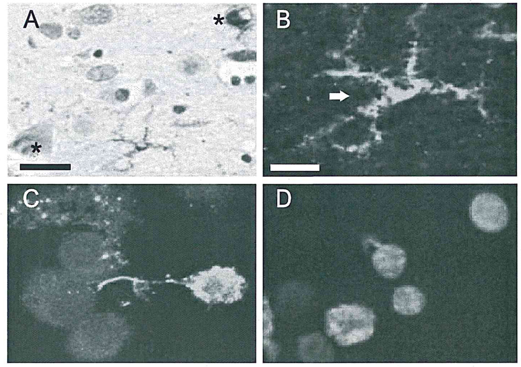
Microglia take up DNA fragments in Alzheimer brain and in vitro. (A and B) TUNEL positivity in cytoplasm (arrow), but not nucleus, in microglia, and in nuclei (asterisk), but not cytoplasm, of neurons in Alzheimer temporal lobe. (A) and (B) are from the same field at different magnifications (A, bar = 25 µm; B, bar= 10 µm). (C) Confocal microscopy demonstrates fluorescein-labeled fDNA within the cytoplasm of N9 microglia. (D) TUNEL-positive fDNA was seen in N9 microglia exposed to unlabeled fDNA, confirming microglial phagocytosis of fDNA.
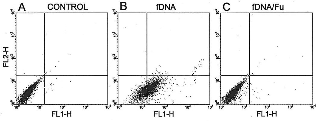
Microglial uptake of DNA fragments is mediated by a scavenger receptor. (A) Untreated sister microglial cultures. (B) fDNA treatment of N9 microglia. (C) fDNA treatment of N9 in the presence of fucoidan (Fu), a general inhibitor of scavenger receptors analyzed by flow cytometry.
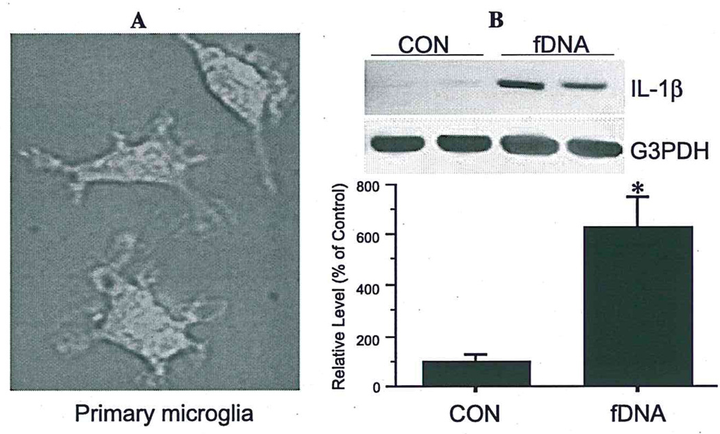
Microglia are activated by internalization of DNA fragments. Fluorescein-labeled fDNA uptake (A) significantly increases IL-1β mRNA expression in primary rat microglia (B). Values are expressed as mean ± S.E.M. of four samples (*p< 0.05).
References
-
- Barger SW, Basile AS. Activation of microglia by secreted amyloid precursor protein evokes release of glutamate by cystine exchange and attenuates synaptic function. J. Neurochem. 2001;76:846–854. - PubMed
-
- Barger SW, Harmon AD. Microglial activation by Alzheimer amyloid precursor protein and modulation by apolipoprotein E. Nature. 1997;388:878–881. - PubMed
-
- Corradin SB, Mauel J, Donini SD, Quattrocchi E, Ricciardi-Castagnoli P. Inducible nitric oxide synthase activity of cloned murine microglial cells. Glia. 1993;7:255–262. - PubMed
Publication types
MeSH terms
Substances
Grants and funding
LinkOut - more resources
Full Text Sources
Other Literature Sources
