

Comparative Study
doi: 10.1016/s0969-8051(03)00119-7.
Imaging recognition of multidrug resistance in human breast tumors using 99mTc-labeled monocationic agents and a high-resolution stationary SPECT system
Affiliations
- PMID: 14741570
- PMCID: PMC3062994
- DOI: 10.1016/s0969-8051(03)00119-7
Item in Clipboard
Comparative Study
Imaging recognition of multidrug resistance in human breast tumors using 99mTc-labeled monocationic agents and a high-resolution stationary SPECT system
Nucl Med Biol.
2004 Jan.
Abstract
Imaging recognition of multidrug-resistance by 99mTc-labeled sestamibi, tetrofosmin and furifosmin in mice bearing human breast tumors was evaluated using a high-resolution SPECT, FASTSPECT. Imaging results showed that the washout rates in drug-resistant MCF7/D40 tumors were significantly greater than that in drug-sensitive MCF7/S tumors. Furifosmin exhibited greater washout from both MCF7/S and MCF7/D40 than sestamibi, while tetrofosmin washout was greater than sestamibi in MCF7/D40 only. Feasibility of the monocationic agents for characterizing MDR expression was well clarified with FASTSPECT imaging.
Figures

Expression of Pgp in MCF7/S and MCF7/D40 human breast tumors from 3 representative mice in each group (99mTc-sestamibi, 99mTc-tetrofosmin and 99mTc-furifosmin), as determined by Western blots of plasma membrane preparations with mAb C219. The arrow identifies Pgp at 170kDa.

Three-dimensional representation (A) of a reconstructed FASTSPECT data set of the right thigh with subcutaneous MCF7/S tumor (arrow) in a mouse (B) 10-min after injection with 99mTc-tetrofosmin. C & D are two selected coronal slices. The tumor (arrow) was visualized on slice D.
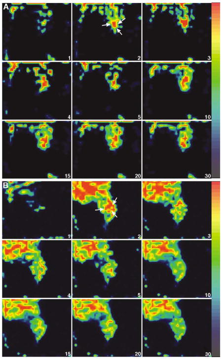
Representative FASTSPECT dynamic images (selected coronal slices) from two mice with MCF7/S breast tumor (A) and MCF7/D40 tumor (B) using 99mTc-sestamibi. The time after injection is shown in the lower right corner of each image. The MCF7/S tumor (arrow) was visualized about 2 min post-injection and stayed well-defined for at least 30 min. The MCF7/D40 tumor (arrow) was visible for only about 2–3 min post-injection. The tumor weight was 0.084 g (MCF7/S) and 0.15 g (MCF7/D40). The ratio of tumor/muscle was 1.45 (MCF7/S) and 0.72 (MCF7/D40) determined by biodistribution analysis at the end of imaging session.

Dynamic images from two mice with MCF7/S breast tumor (A) and MCF7/D40 tumor (B) using 99mTc-tetrofosmin (transaxial slices). The MCF7/S tumor (arrow) was visible clearly from 2 min to 30 min consistently. The MCF7/D40 tumor (arrow) was well identified at 2 min post-injection. After 3 min, the radioactivity in the tumor quickly dropped to the background level. The tumor weight was 0.09 g (MCF7/S) and 0.04 g (MCF7/D40). The ratio of tumor/muscle was 1.31 (MCF7/S) and 0.59 (MCF7/D40), respectively.
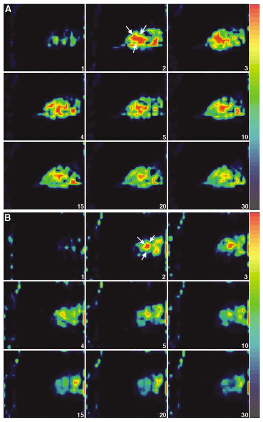
99mTc-furifosmin dynamic images (sagittal slices) from two mice with MCF7/S breast tumor (A) and MCF7/D40 tumor (B). The MCF7/S tumor (arrow) became visible 2–5 min and remained detectable until 30 min post-injection. The MCF7/D40 tumor (arrow) was visualized 2–5 min post-injection. The tumor weight was 0.11 g (MCF7/S) and 0.07 g (MCF7/D40). The ratio of tumor/muscle was 0.93 (MCF7/S) and 0.41 (MCF7/D40), respectively.
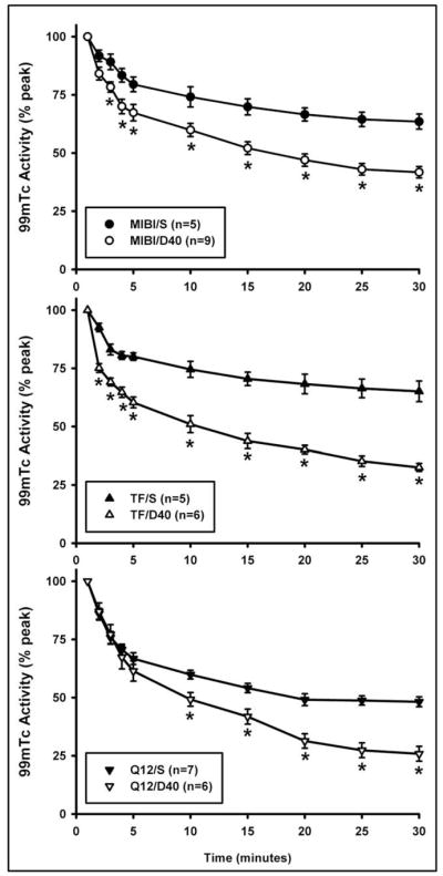
99mTc-sestamibi (MIBI), 99mTc-tetrofosmin (TF) and 99mTc-furifosmin (Q12) clearance curves from MCF7/S and MCF7/D40 breast tumors. The radioactivity is plotted as percent of the peak activity in the tumor. S = MCF7/S; D40 = MCF7/D40.

99mTc-sestamibi, 99mTc-tetrofosmin and 99mTc-furifosmin fractional washout (%) from MCF7/S and MCF7/D40 tumors. * p<0.05 compared to 99mTc-sestamibi; + p<0.05 compared to 99mTc-tetrofosmin.
References
-
- Pastan I, Gottesman M. Multiple-drug resistance in human cancer. N Engl J Med. 1987;316:1388–93. - PubMed
-
- Woodhouse JR, Ferry DR. The genetic basis of resistance to cancer chemotherapy. Ann Med. 1995;27:157–67. - PubMed
-
- Gottesman MM, Pastan I. The multidrug transporter, a double-edged sword. J Biol Chem. 1988;263:12163–6. - PubMed
-
- Bellamy WT. P-glycoproteins and multidrug resistance. Annu Rev Pharmacol Toxicol. 1996;36:161–83. - PubMed
-
- Gottesman MM, Pastan I. Biochemistry of multidrug resistance mediated by the multidrug transporter. Annu Rev Biochem. 1993;62:385–427. - PubMed
Publication types
MeSH terms
Substances
Grants and funding
LinkOut - more resources
Full Text Sources
Medical
Research Materials
