. 2004 Feb;48(2):546-55.
doi: 10.1128/AAC.48.2.546-555.2004.

Subinhibitory concentrations of linezolid reduce Staphylococcus aureus virulence factor expression

Affiliations

Subinhibitory concentrations of linezolid reduce Staphylococcus aureus virulence factor expression

Katussevani Bernardo et al. Antimicrob Agents Chemother. 2004 Feb.

Abstract

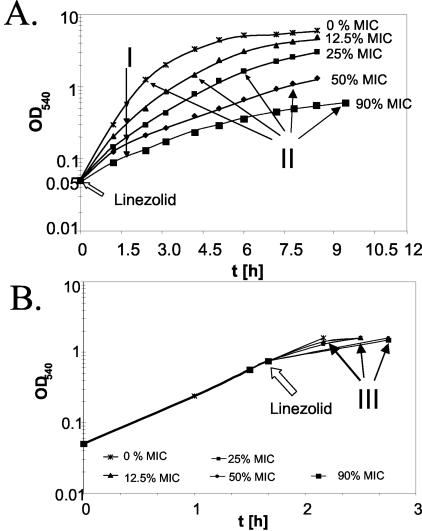
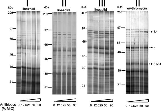
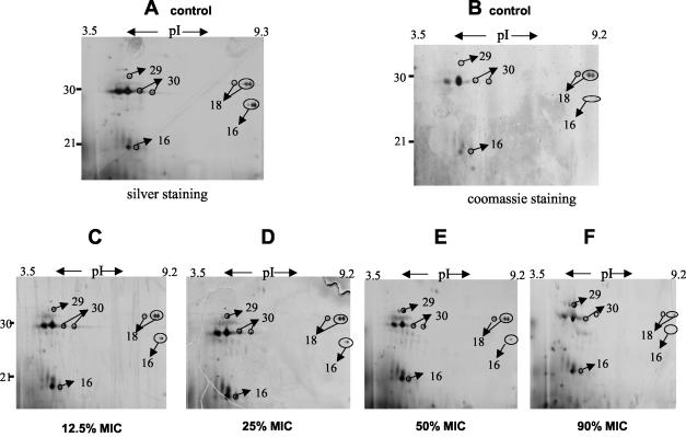
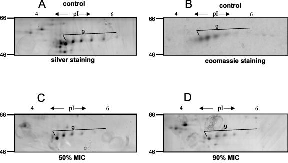
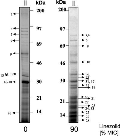
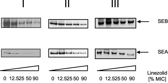
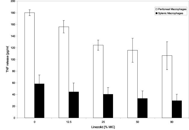
The influence of the antibiotic linezolid on the secretion of exotoxins by Staphylococcus aureus was analyzed by sodium dodecyl sulfate-polyacrylamide gel electrophoresis combined with matrix-assisted laser desorption ionization-time of flight mass spectrometry and Western blot analysis. S. aureus suspensions were treated with grading subinhibitory concentrations of linezolid (12.5, 25, 50, and 90% of MIC) at different stages of bacterial growth (i.e., an optical density at 540 nm [OD(540)] of 0.05 or 0.8). When added to S. aureus cultures at an OD(540) of 0.05, linezolid reduced in a dose-dependent manner the secretion of specific virulence factors, including staphylococcal enterotoxin A (SEA) and SEB, bifunctional autolysin, autolysin, protein A, and alpha- and beta-hemolysins. In contrast, other presumably nontoxic exoproteins remained unchanged or even accumulated in supernatants in the presence of linezolid at a 90% MIC. Similarly, when added at OD(540) of 0.8, that is, after quorum sensing, linezolid reduced the release of virulence factors, whereas the relative abundance of nontoxic exoproteins such as triacylglycerol lipase, glycerol ester hydrolase, DnaK, or translation elongation factor EF-Tu was found to be increased. Consistently, linezolid reduced in a dose-dependent manner the tumor necrosis factor-inducing activity secreted by S. aureus into the culture supernatants. The results of our study suggest that the expression of virulence factors in S. aureus is especially sensitive to the inhibition of protein synthesis by linezolid, which should be an advantage in the treatment of infections with toxin-producing S. aureus.


Figures

FIG. 1.
FIG. 1.
Effect of linezolid on growth of S. aureus. S. aureus strain ATCC 29213 was grown at 37°C. At indicated time points, the OD540 was measured. (A) Bacteria were grown in the presence of grading concentrations of linezolid from the start of growth (OD540 ∼0.05) and harvested after 2 h (I) or after they reached an OD540 of ∼1.0 (II). (B) Bacteria were grown first to an OD540 of 0.8; thereafter, growth was continued in the presence of linezolid with increasing concentrations and harvested at an OD540 of ∼1.0 (III). Arrows indicate the time point of harvest.
FIG. 2.
FIG. 2.
SDS-PAGE of exotoxin production by S. aureus in the presence of antibiotics. Culture supernatants of S. aureus treated with different concentrations of linezolid or erythromycin were analyzed by SDS-PAGE as described in Materials and Methods. Protein bands were visualized by silver staining (sample of 10 μg of protein). Panels I to III correspond to the conditions described in the legend to Fig. 1. The arrows indicate the protein bands subjected to MALDI-TOF/MS and/or LC-MS analysis.
FIG. 3.
FIG. 3.
Two-dimensional patterns of exoproteins in the presence of graded concentrations of linezolid. (Upper panels [A to F]) Partial view (pI 3 to 10 and 10 to 40 kDa); (lower panels [A to D]) partial view (pI 3.2 to 6.9 and 46 to 66 kDa). S. aureus ATCC 29213 was grown at 37°C with increasing concentrations of linezolid from the start of growth (OD540 ∼0.05) and harvested at an OD540 of ∼1.0. Supernatants of S. aureus cultures were subjected to two-dimensional gel electrophoresis. Lettered panels: A and B, untreated; C to F treated with linezolid at 12.5, 25, 50, and 90% MICs, respectively. Protein spots were visualized by silver staining (A and C to F [sample of 100 μg of protein]) or Coomassie blue stain (B [sample of 500 μg of protein]). The arrows indicate the protein bands subjected to MALDI-TOF/MS and/or LC-MS analysis.
FIG. 3.
FIG. 3.
Two-dimensional patterns of exoproteins in the presence of graded concentrations of linezolid. (Upper panels [A to F]) Partial view (pI 3 to 10 and 10 to 40 kDa); (lower panels [A to D]) partial view (pI 3.2 to 6.9 and 46 to 66 kDa). S. aureus ATCC 29213 was grown at 37°C with increasing concentrations of linezolid from the start of growth (OD540 ∼0.05) and harvested at an OD540 of ∼1.0. Supernatants of S. aureus cultures were subjected to two-dimensional gel electrophoresis. Lettered panels: A and B, untreated; C to F treated with linezolid at 12.5, 25, 50, and 90% MICs, respectively. Protein spots were visualized by silver staining (A and C to F [sample of 100 μg of protein]) or Coomassie blue stain (B [sample of 500 μg of protein]). The arrows indicate the protein bands subjected to MALDI-TOF/MS and/or LC-MS analysis.
FIG. 4.
FIG. 4.
Identification of exoproteins of S. aureus treated with linezolid. Supernatants of S. aureus cultures left untreated (left) or treated with linezolid at 90% MIC from the start of growth (OD540 ∼0.05) (right) were harvested at an OD540 of ∼0.8. Protein bands were visualized by coomassie staining. The arrows indicate the protein bands subjected to MALDI-TOF/MS or LC-MS/MS analysis.
FIG. 5.
FIG. 5.
Western blot analysis of exotoxins of S. aureus treated with linezolid. S. aureus ATCC 29213 was grown at 37°C and treated with linezolid at various growth phases. Panels I to III correspond to the conditions described in the legend to Fig. 1. Supernatants were subjected to SDS-PAGE. After transfer to nitrocellulose, proteins were stained specifically with the indicated antibodies against SEA and SEB. A horseradish peroxidase-conjugated goat anti-rabbit antibody was used as second reagent and visualized by using an enhanced chemiluminescence detection kit (Amersham-Pharmacia).
FIG. 6.
FIG. 6.
TNF release by macrophages stimulated with supernatants of S. aureus. S. aureus strains were cultured in DMEM and treated with the indicated concentrations of linezolid as described in the text. Secreted proteins present in the supernatant were collected, followed by incubation for 20 h at 37°C with mouse splenic macrophages or peritoneal macrophages. The TNF levels were measured by ELISA. Linezolid itself (1× MIC and 2× MIC) did not induce TNF (data not shown).

References

    1. Arvidson, A., and K. Tegmark. 2001. Regulation of virulence determinants in Staphylococcus aureus. Int. J. Med. Microbiol. 291:159-170. - PubMed
    1. Bassler, B. L. 1999. How bacteria talk to each other: regulation of gene expression by quorum sensing. Curr. Opin. Microbiol. 2:582-587. - PubMed
    1. Bernardo, K., S. Fleer, N. Pakulat, O. Krut, F. Hünger, and M. Krönke. 2002. Identification of Staphylococcus aureus exotoxins by combined SDS gel electrophoresis and MALDI-TOF-MS. Proteomics 2:740-746. - PubMed
    1. Bernstein, J. A., A. B. Khodursky, P. H. Lin, S. Lin-Chao, and S. N. Cohen. 2002. Global analysis of mRNA decay and abundance in Escherichia coli at single-gene resolution using two-color fluorescent DNA microarrays. Proc. Natl. Acad. Sci. USA 99:9697-9702. - PMC - PubMed
    1. Bjorklind, A., and A. Arvidson. 1980. Mutants of Staphylococcus aureus affected in the regulation of exoprotein synthesis. FEMS Microbiol. Lett. 7:203-206.

Publication types

MeSH terms

LinkOut - more resources