. 2004 Feb;164(2):577-88.
doi: 10.1016/S0002-9440(10)63147-1.

Basal cells are a multipotent progenitor capable of renewing the bronchial epithelium

Affiliations

Basal cells are a multipotent progenitor capable of renewing the bronchial epithelium

Kyung U Hong et al. Am J Pathol. 2004 Feb.

Abstract

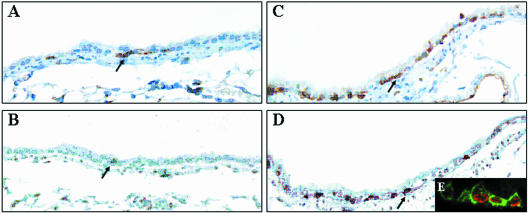
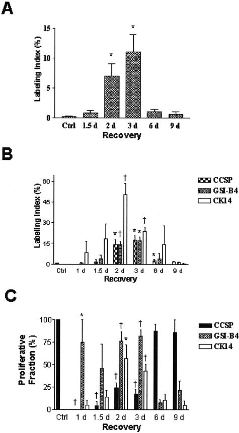
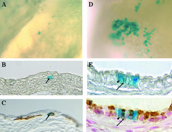
Commitment of the pulmonary epithelium to bronchial and bronchiolar airway lineages occurs during the transition from pseudoglandular to cannalicular phases of lung development, suggesting that regional differences exist with respect to the identity of stem and progenitor cells that contribute to epithelial maintenance in adulthood. We previously defined a critical role for Clara cell secretory protein-expressing (CE) cells in renewal of bronchiolar airway epithelium following injury. Even though CE cells are also the principal progenitor for maintenance of the bronchial airway epithelium, CE cell injury is resolved through a mechanism involving recruitment of a second progenitor cell population that we now identify as a GSI-B(4) reactive, cytokeratin-14-expressing basal cell. These cells exhibit multipotent differentiation capacity as assessed by analysis of cellular phenotype within clones of LacZ-tagged cells. Clones were derived from K14-expressing cells tagged in a cell-type-specific fashion by ligand-regulable Cre recombinase-mediated genomic rearrangement of the ROSA26 recombination substrate allele. We conclude that basal cells represent an alternative multipotent progenitor cell population of bronchial airways and that progenitor cell selection is dictated by the type of airway injury.


Figures

Figure 1
Figure 1
Proliferation and hyperplasia of basal cells in the bronchial epithelium of GCV-treated CCtk transgenic mice. CCtk mice were acutely exposed to vehicle (A and B) or 10 mg GCV (C and D), continuously exposed to BrdU during days 2 to 8 of the recovery period, and sacrificed on recovery day 10. Basal cells were detected by GSI-B4 lectin histochemistry (A and C) and S-phase cells were detected on adjacent serial sections by immunohistochemical detection of BrdU (C and D). Lectin and immune complexes were detected with diaminobenzidine (brown stain). Original magnification, ×400. Tissue sections from animals treated with GCV and recovered 3 days were analyzed by dual-immunofluorescence detection of GSI-B4 (green fluorescence) and BrdU (red fluorescence) and were analyzed by confocal microscopy (E). Original magnification, ×600.
Figure 2
Figure 2
Cellular composition of the normal and regenerating bronchial epithelium. Lung tissue sections from untreated (A, E, and I) and naphthalene-treated mice that had recovered 3 days (B, F, and J), 6 days (C, G, and K), or 9 days (D, H, and L) were immunostained for CCSP (A to D), GSI-B4 (E to H), or K14 (I to L). Tissue was counterstained with hematoxylin and images of the lobar bronchus collected using standard light microscopy. Original magnification, ×400. Cellular composition (M) within the bronchial epithelium of control (Ctrl) or naphthalene-treated mice that had been recovered 1.5 to 9 days (d) was defined as the number of immunoreactive cells in each category divided by the length on the BM in millimeters. The mean ± SEM (n = 4 to 5) is presented for each group. *, P < 0.05 relative to values for untreated control mice.
Figure 3
Figure 3
Bronchial proliferation following naphthalene-induced Clara cell depletion. Corn oil (A, F, K) or naphthalene-treated mice were recovered 1.5 days (B, G, L), 3 days (C, H, M), 6 days (D, I, N), or 9 days (E, J, O) and injected with BrdU 2 hours before sacrifice. Adjacent serial sections from each experimental group were analyzed by dual immunofluorescence for CCSP (A to E, red) and BrdU (green), GSI-B4 binding site (F to J, green) and BrdU (red), or K14 (K to O, green) and BrdU (red).
Figure 4
Figure 4
Labeling index and proliferation fraction of the bronchial epithelium. The total labeling index for the bronchial epithelium (A) was defined as the percentage of BrdU-labeled nuclei/total number of nuclei. The labeling index for each cell type (B) was defined as the percentage of dual-positive CCSP/BrdU, GSI-B4/BrdU, or K14/BrdU cells divided by the total number of cells in each category. The proliferative fraction (C) was defined as the percentage of BrdU-labeled cells of each cell type divided by the total number of BrdU-labeled nuclei. Each value was determined for control (Ctrl) and naphthalene-treated mice that had been recovered 1.5 to 9 days (d) and is reported as the mean ± SEM (n = 4 to 5 for each group). *, P < 0.05 and †, P < 0.01 relative to values in untreated control mice.
Figure 5
Figure 5
CCSP/K14 dual-positive cells. The occurrence of bronchial cells that co-express CCSP (A, green) and K14 (B, red) was determined by dual immunofluorescence and confocal microscopy. The single-color images were merged (C) and used to determine the number of dual-positive cells per millimeter of BM in control (Ctrl) and naphthalene-treated mice following 1.5 to 9 days (d) of recovery (D). The images presented in A to C are from an animal that had been recovered 6 days. Original magnification, ×600. The histograms represent the mean ± SEM (n = 4 to 5). *, P < 0.05.
Figure 6
Figure 6
Lineage of cytokeratin-14 expressing cells of the bronchial epithelium. K14-cre-ERt/RS bitransgenic mice were treated with naphthalene on day 0 and with Tamoxifen on recovery days 2 to 4. β-gal-expressing cells within the bronchial epithelium (blue-stained cells) were detected by wholemount histochemistry on recovery days 4 (A to C) and 20 (D to F). Tissue was initially imaged as a wholemount (A and D) and subsequently embedded, sectioned, and re-imaged (B and E). Sections containing β-gal-expressing cells were analyzed by immunohistochemistry for expression of K14 (C) or CCSP (F). Arrows in B and C indicate a β-gal/K14 dual-positive cell. Arrowheads in E and F indicate a β-gal-positive ciliated cell and the arrows indicate a β-gal/CCSP dual-positive cell.

References

    1. Shannon JM. Induction of alveolar type II cell differentiation in fetal tracheal epithelium by grafted distal lung mesenchyme. Dev Biol. 1994;166:600–614. - PubMed
    1. Shannon JM, Nielsen LD, Gebb SA, Randell SH. Mesenchyme specifies epithelial differentiation in reciprocal recombinants of embryonic lung and trachea. Dev Dyn. 1998;212:482–494. - PubMed
    1. Jeffery PK, Reid L. New observations of rat airway epithelium: a quantitative and electron microscopic study. J Anat. 1975;120:295–320. - PMC - PubMed
    1. Evans MJ, Moller PC. Biology of airway basal cells. Exp Lung Res. 1991;17:513–531. - PubMed
    1. Inayama Y, Hook GER, Brody AR, Cameron GS, Jetten AM, Gilmore LB, Gray T, Nettesheim P. The differentiation potential of tracheal basal cells. Lab Invest. 1988;58:706–717. - PubMed

Publication types

MeSH terms