. 2004 Feb;164(2):623-34.
doi: 10.1016/S0002-9440(10)63151-3.

Collapse and restoration of MHC class-I-dependent immune privilege: exploiting the human hair follicle as a model

Affiliations

Collapse and restoration of MHC class-I-dependent immune privilege: exploiting the human hair follicle as a model

Taisuke Ito et al. Am J Pathol. 2004 Feb.

Abstract

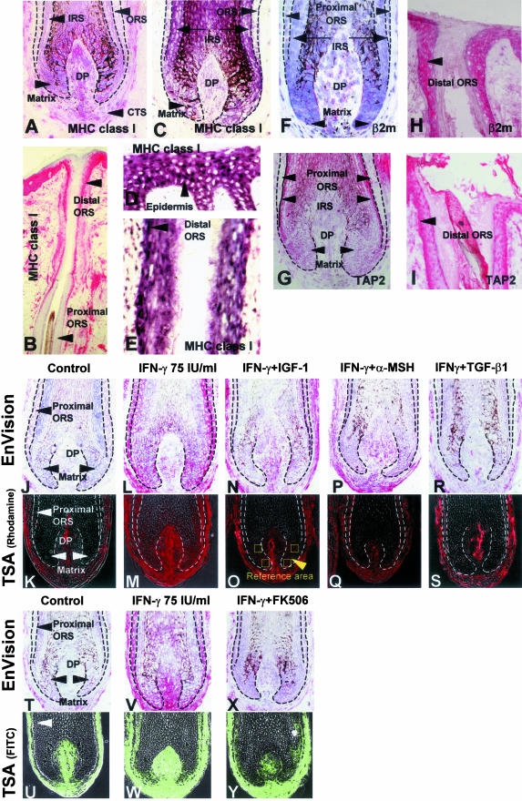
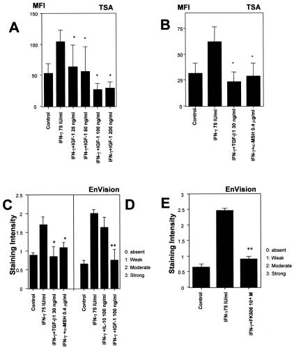
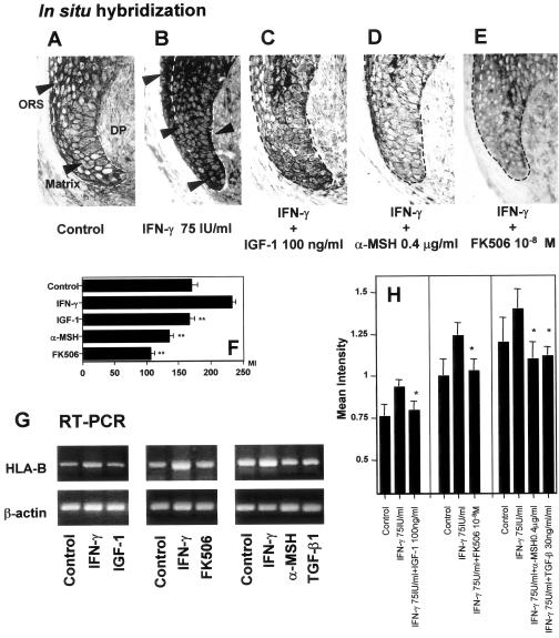
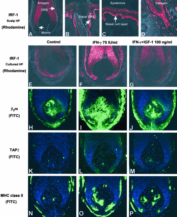
The collapse of major histocompatibility complex (MHC) class-I-dependent immune privilege can lead to autoimmune disease or fetal rejection. Pragmatic and instructive models are needed to clarify the as yet obscure controls of MHC class I down-regulation in situ, to dissect the principles of immune privilege generation, maintenance, and collapse as well as to develop more effective strategies for immune privilege restoration. Here, we propose that human scalp hair follicles, which are abundantly available and easily studied, are ideally suited for this purpose: interferon-gamma induces ectopic MHC class I expression in the constitutively MHC class-I-negative hair matrix epithelium of organ-cultured anagen hair bulbs, likely via interferon regulatory factor-1, along with up-regulation of the MHC class I pathway molecules beta(2)microglobulin and transporter associated with antigen processing (TAP-2). In the first report to identify natural immunomodulators capable of down-regulating MHC class I expression in situ in a normal, neuroectoderm-derived human tissue, we show that ectopic MHC class I expression in human anagen hair bulbs can be normalized by treatment with alpha-MSH, IGF-1, or TGF-beta1, all of which are locally generated, as well as by FK506. These agents are promising candidates for immune privilege restoration and for suppressing MHC class I expression where this is clinically desired (eg, in alopecia areata, multiple sclerosis, autoimmune uveitis, mumps orchitis, and fetal or allograft rejection).


Figures

Figure 1
Figure 1
A to I: Anagen hair bulbs express very low epithelial MHC class I and its pathway molecules in normal human scalp skin sections. A: Extra-sensitive APAAP-based immunohistology techniques (EnVision) show very low or absent MHC class-I-like immunoreactivity in the hair matrix, inner root sheath (IRS), and proximal ORS. Dermal papilla cells show marked variation in MHC class I immunoreactivity between individuals. Some dermal papilla cells have strong immunoreactivity for MHC class I and others have only weak immunoreactivity. B: The epidermis, distal ORS, and the connective tissue sheath of anagen VI scalp hair follicle have high MHC class-I-like immunoreactivity. In the ORS, MHC class I immunoreactivity declines from distal to proximal. C: On in situ hybridization technique, the proximal ORS and matrix keratinocytes show very little detectable HLA-B gene expression, supporting the MHC class I protein expression data (A). D and E: Note the strongly positive HLA-B mRNA signals in the epidermis (human scalp skin) and the distal ORS. F: Proximal ORS and hair matrix keratinocytes show low or absent expression level of β2microglobulin immunoreactivity similar to the expression of MHC class I. In comparison with proximal ORS and hair matrix keratinocytes, dermal papilla cells express moderate expression of β2microglobulin. G: Proximal ORS, hair matrix, and dermal papilla cells show low expression of TAP2 immunoreactivity. H: Distal ORS and epidermis show high levels of β2microglobulin immunoreactivity. I: Distal ORS shows high level of TAP2 immunoreactivity. J to Y: IGF-1, α-MSH, TGF-β1, and FK506 down-regulate IFN-γ-induced ectopic MHC class I protein expression in vitro. J and K: EnVision (J) and Tyramide signal amplification (TSA) (K) show the low expression of MHC class I in the vehicle-treated control follicle matrix and proximal ORS in cultured human hair follicles. L and M: 75 IU/m IFN-γ induces the ectopic MHC class I expression in human hair matrix and proximal ORS. N to S: 100 ng/ml IGF-1 (N and O), 0.4 μg/ml α-MSH (P and Q), and 30 ng/ml TGF-β1 (R and S) down-regulate the IFN-γ-induced ectopic MHC class I expression in human hair matrix and proximal ORS. T to Y: In comparison with vehicle-treated control hair follicles (T and U), 75 IU/ml IFN-γ-treated human hair matrix has a strong MHC class I immunoreactivity (V and W), which is down-regulated by 1 × 10⁻⁸ FK506 (X and Y) in vitro. The mean fluorescence intensity (MFI) was measured at four previously defined reference areas in the hair matrix of anagen hair bulbs (N and O) by NIH image, and the average MFI was calculated [n = 60 hair follicles (HFs) from 3 patients/group]. β2m, β2microglobulin; CTS, connective tissue sheath; DP, dermal papilla.
Figure 2
Figure 2
IGF-1, TGF-β, α-MSH, and FK506 can down-regulate IFN-γ-induced ectopic MHC class I expression in hair matrix. A: IGF-1 (25 to 200 ng/ml) down-regulates the fluorescent mean intensity of MHC class I in hair matrix by dose dependent manner. B: TSA, which is high-sensitive immunohistochemical technique, revealed that 30 ng/ml TGF-β1 and 0.4 μg/ml α-MSH significantly down-regulate the mean intensity of MHC class I in the hair matrix. C and D: EnVision also shows that 100 ng/ml IGF-1, 30 ng/ml TGF-β1, 0.4 μg/ml α-MSH significantly down-regulate the staining intensity of MHC class I in the hair matrix. D: IL-10 fails to down-regulate the IFN-γ-induced ectopic MHC class I expression. E: The expression of MHC class I was significantly down-regulated in hair matrix treated by 10−8 M FK506. The MFI of MHC class I expression was measured at four previously defined reference areas in the hair matrix of anagen hair bulbs by NIH image, and the average MFI was calculated (n = 60 HFs from 3 patients/group). Staining intensity of MHC class I-immunoreactivity was scored as follows: −, absent; +, weak; ++, moderate; +++, strong (n = 60 HFs from 3 patients/each group), indicated as mean values ± SEM (n = 60 HF/group from 3 patients). *, P < 0.05 versus IFN-γ-treated hair follicles; **, P < 0.01 versus IFN-γ-treated hair follicles.
Figure 3
Figure 3
HLA-B gene expression is down-regulated by IGF-1, α-MSH, TGF-β, and FK506. A: In situ hybridization revealed that HLA-B mRNA expression was very low in control hair matrix and proximal hair ORS which is same as the result shown in Figure 1. B: 75 U/ml IFN-γ strongly up-regulated HLA-B mRNA expression in matrix. C: 100 ng/ml IGF-1 repressed the number of ectopic HLA-B mRNA-like signals. D: 0.4 μg/ml α-MSH repressed the number of ectopic HLA-B mRNA-like signals. E: 1 × 10⁻⁸ M FK506 repressed the number of ectopic HLA-B mRNA-like signals. F: The mean intensity of mRNA expression by in situ hybridization was evaluated by NIH image in matrix and distal ORS. G and H: Semiquantitative RT-PCR supports the result by immunohistochemistry that shows IGF-1, FK506, α-MSH, and TGF-β1 inhibits 75 IU/ml IFN-γ-induced ectopic HLA-B mRNA transcription. The mean intensity of each amplified signals were measured and compared with that of β-actin using NIH image software. The level of mean intensity ratio was down-modulated in HF treated with 100 ng/ml IGF-1, 1 × 10⁻⁸M FK506, 0.4 μg/ml, and 30 ng/ml TGF-β1. *, P < 0.05 versus IFN-γ-treated hair follicles; ** P < 0.01 versus IFN-γ-treated HFs.
Figure 4
Figure 4
A: HF matrix has absent or low IRF-1-like-immunoreactivity in anagen hair bulb. B: In anagen hair follicle, IRF-1 immunoreactivity is declined from epidermis to distal ORS. C: Basal layer epidermis shows IRF-1 strong immunoreactivity in human scalp skin. D: Very prominent immunoreactivity throughout the epithelium of catagen follicles. E: Control HF shows low immunoreactivity of IRF-1. F and G: 75 IU/ml IFN-γ up-regulated IRF-1 immunoreactivity in matrix and proximal ORS (F), which is restored by 100 ng/ml IGF-1 (G). H to P: IFN-γ-induced ectopic MHC class I pathway molecules and MHC class II immunoreactivity was down-regulated by IGF-1. H: TSA revealed that control HF has very low immunoreactivity of β2microglobulin like MHC class I expression. I and J: 75 IU/ml IFN-γ up-regulate β2microglobulin immunoreactivity in hair follicle matrix, and down-regulated by 100 ng/ml IGF-1. K to M: IFN-γ-induced ectopic TAP2 immunoreactivity was also down-regulated by 100 ng/ml IGF-1. IGF-1 also down-regulates IFN-γ- induced MHC class II expression. N: The expression of MHC class II in control HFs were very low in matrix and proximal ORS. O to P: Control HF shows a few MHC class-II-positive cells in the connective tissue sheath; 75 IU/ml IFN-γ prominently induced MHC class II expression not only in the connective tissue sheath but also in the proximal ORS (keratinocytes), which could be down-regulated again by 100 ng/ml IGF-1 (P).

References

    1. Head JR, Billingham RE. Immunologically privileged sites in transplantation immunology and oncology. Perspect Biol Med. 1985;29:115–131. - PubMed
    1. Streilein JW. Immune privilege as the result of local tissue barriers and immunosuppressive microenvironments. Curr Opin Immunol. 1993;5:428–432. - PubMed
    1. Niederkorn JY. Immunology and immunomodulation of corneal transplantation. Int Rev Immunol. 2002;21:173–196. - PubMed
    1. Mellor AL, Munn DH. Immunology at the maternal-fetal interface: lessons for T cell tolerance and suppression. Annu Rev Immunol. 2000;18:367–391. - PubMed
    1. Erlebacher A. Why isn’t the fetus rejected? Curr Opin Immunol. 2001;13:590–593. - PubMed

Publication types

MeSH terms