

The impact of gender on progression of renal disease: potential role of estrogen-mediated vascular endothelial growth factor regulation and vascular protection
- PMID: 14742271
- PMCID: PMC1602256
- DOI: 10.1016/S0002-9440(10)63155-0
The impact of gender on progression of renal disease: potential role of estrogen-mediated vascular endothelial growth factor regulation and vascular protection
Abstract
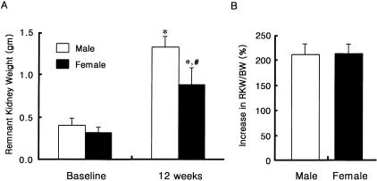
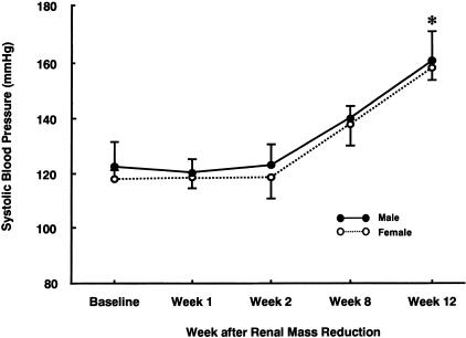
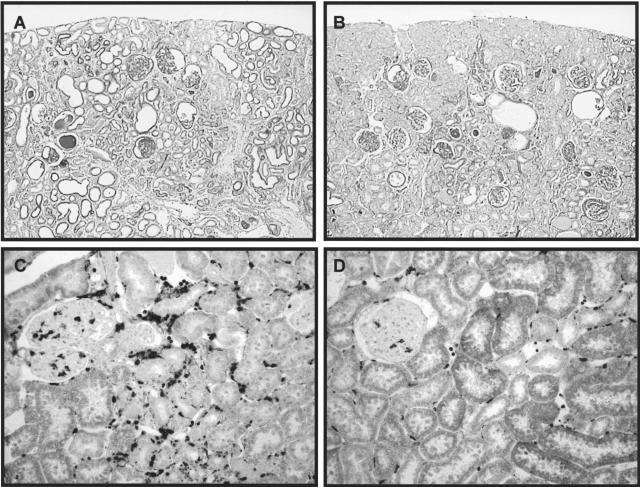
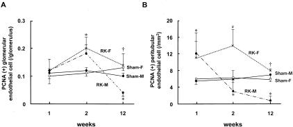
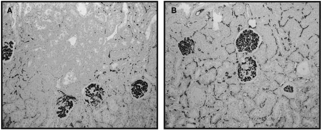
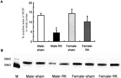
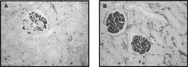
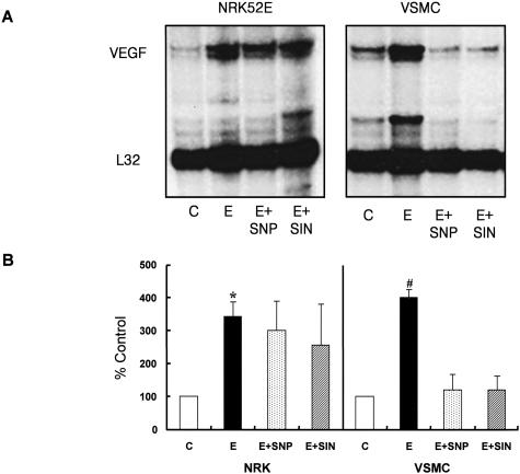
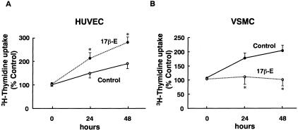
Male gender is associated with a more rapid progression of renal disease independent of blood pressure, dietary protein intake, or serum lipid levels. Recently, we reported a key role for the intrarenal vasculature in progressive renal disease (Kang D-H, Kanellis J, Hugo C, Truong L, Anderson S, Kerjaschki D, Schreiner GF, Johnson RJ: Role of endothelium in progressive renal disease. J Am Soc Nephrol 2002, 13:806-816). We hypothesized that estrogen-mediated preservation of the renal vasculature could account for the better renal outcome in female rats. We analyzed micro- and macrovascular changes in the 5/6 remnant kidney (RK) models both in male (n = 24) and female (n = 24) Sprague-Dawley rats up to 12 weeks after renal mass reduction. At 12 weeks, male and female RK rats had equivalent blood pressure, glomerular tuft area, and RK/body weight, but male rats showed worse renal function, proteinuria, glomerulosclerosis (%), and tubulointerstitial fibrosis. At 12 weeks peritubular capillary (PTC) EC proliferation and PTC density were higher in female RK rats whereas macrovascular changes in preglomerular vessels (smooth muscle cell proliferation, medial wall thickening, and adventitial fibrosis) were less prominent. The expression of vascular endothelial growth factor (VEGF) and VEGF type 2 receptor (flk-1) in renal cortex assessed by immunostaining were higher in female RK rats. To dissect the mechanism of sex hormone-induced vascular remodeling and VEGF regulation, we investigated the in vitro effect of 17 beta-estradiol (17 beta E, 10 nmol/L) on proliferation and VEGF expression of renal tubular cells (rat proximal tubular cells), vascular smooth muscle cells (VSMCs), and human umbilical vein endothelial cells (HUVECs). 17 beta E directly stimulated the proliferation of HUVECs, whereas it inhibited serum-induced proliferation of VSMCs. 17 beta E stimulated VEGF mRNA expression both in renal tubular cells and VSMCs. However, when cells were pretreated with a nitric oxide donor to simulate the in vivo condition, 17 beta E inhibited VEGF mRNA expression and protein release in VSMCs. In conclusion, female RK rats developed less glomerulosclerosis and renal failure compared to male RK rats in association with greater preservation of PTC and less preglomerular arteriopathy. Estrogen stimulated basal VEGF expression in renal tubular cells. We propose that estrogen may protect female rats in progressive renal disease by stimulating VEGF expression and maintaining a healthy intrarenal vasculature.
Figures
References
-
- Silbiger SR, Neugarten J. The impact of gender on the progression of chronic renal disease. Am J Kidney Dis. 1995;25:515–533. - PubMed
-
- Neugarten J, Acharya A, Silbiger SR. Effect of gender on the progression of nondiabetic renal disease: a meta-analysis. J Am Soc Nephrol. 2000;11:319–329. - PubMed
-
- Dubey RK, Jackson EK. Estrogen-induced cardiorenal protection: potential cellular, biochemical, and molecular mechanisms. Am J Physiol. 2001;280:F365–F388. - PubMed
-
- Kang DH, Anderson S, Kim YG, Mazzalli M, Suga S, Jefferson JA, Gordon KL, Oyama TT, Hughes J, Hugo C, Kerjaschki D, Schreiner GF, Johnson RJ. Impaired angiogenesis in the aging kidney: potential role of VEGF and TSP-1 in renal disease. Am J Kidney Dis. 2001;37:601–611. - PubMed
-
- Kang DH, Joly AH, Oh SW, Hugo C, Kerjaschki D, Gordon KL, Mazzali M, Jefferson JA, Hughes J, Madsen KM, Schreiner GF, Johnson RJ. Impaired angiogenesis in the remnant kidney model (I): potential role of vascular endothelial growth factor and thrombospondin-1. J Am Soc Nephrol. 2001;12:1434–1447. - PubMed
Publication types
MeSH terms
Substances
Grants and funding
LinkOut - more resources
Full Text Sources
Medical
