

Surfactant protein A modulates the inflammatory response in macrophages during tuberculosis
- PMID: 14742504
- PMCID: PMC321592
- DOI: 10.1128/IAI.72.2.645-650.2004
Surfactant protein A modulates the inflammatory response in macrophages during tuberculosis
Abstract
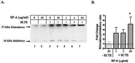
Tuberculosis leads to immune activation and increased human immunodeficiency virus type 1 (HIV-1) replication in the lung. However, in vitro models of mycobacterial infection of human macrophages do not fully reproduce these in vivo observations, suggesting that there are additional host factors. Surfactant protein A (SP-A) is an important mediator of innate immunity in the lung. SP-A levels were assayed in the human lung by using bronchoalveolar lavage (BAL). There was a threefold reduction in SP-A levels during tuberculosis only in the radiographically involved lung segments, and the levels returned to normal after 1 month of treatment. The SP-A levels were inversely correlated with the percentage of neutrophils in BAL fluid, suggesting that low SP-A levels were associated with increased inflammation in the lung. Differentiated THP-1 macrophages were used to test the effect of decreasing SP-A levels on immune function. In the absence of infection with Mycobacterium tuberculosis, SP-A at doses ranging from 5 to 0.01 micro g/ml inhibited both interleukin-6 (IL-6) production and HIV-1 long terminal repeat (LTR) activity. In macrophages infected with M. tuberculosis, SP-A augmented both IL-6 production and HIV-1 LTR activity. To better understand the effect of SP-A, we measured expression of CAAT/enhancer binding protein beta (C/EBPbeta), a transcription factor central to the regulation of IL-6 and the HIV-1 LTR. In macrophages infected with M. tuberculosis, SP-A reduced expression of a dominant negative isoform of C/EBPbeta. These data suggest that SP-A has pleiotropic effects even at the low concentrations found in tuberculosis patients. This protein augments inflammation in the presence of infection and inhibits inflammation in uninfected macrophages, protecting uninvolved lung segments from the deleterious effects of inflammation.
Figures


Similar articles
-
Type I interferon induces inhibitory 16-kD CCAAT/ enhancer binding protein (C/EBP)beta, repressing the HIV-1 long terminal repeat in macrophages: pulmonary tuberculosis alters C/EBP expression, enhancing HIV-1 replication.J Exp Med. 1998 Oct 5;188(7):1255-65. doi: 10.1084/jem.188.7.1255. J Exp Med. 1998. PMID: 9763605 Free PMC article.
-
[Tuberculosis in patients with acquired immune deficiency syndrome].Kekkaku. 2000 Sep;75(9):547-56. Kekkaku. 2000. PMID: 11068371 Review. Japanese.
-
[Molecular pathogenesis in tuberculosis complicated with AIDS].Kekkaku. 2004 Nov;79(11):659-67. Kekkaku. 2004. PMID: 15729891 Review. Japanese.
-
Maximal HIV-1 replication in alveolar macrophages during tuberculosis requires both lymphocyte contact and cytokines.J Exp Med. 2002 Feb 18;195(4):495-505. doi: 10.1084/jem.20011614. J Exp Med. 2002. PMID: 11854362 Free PMC article.
-
Differentiation of monocytes to macrophages switches the Mycobacterium tuberculosis effect on HIV-1 replication from stimulation to inhibition: modulation of interferon response and CCAAT/enhancer binding protein beta expression.J Immunol. 2000 Aug 15;165(4):2028-39. doi: 10.4049/jimmunol.165.4.2028. J Immunol. 2000. PMID: 10925286
Cited by
-
Human Pulmonary Tuberculosis: Understanding the Immune Response in the Bronchoalveolar System.Biomolecules. 2022 Aug 20;12(8):1148. doi: 10.3390/biom12081148. Biomolecules. 2022. PMID: 36009042 Free PMC article. Review.
-
The Importance of First Impressions: Early Events in Mycobacterium tuberculosis Infection Influence Outcome.mBio. 2016 Apr 5;7(2):e00342-16. doi: 10.1128/mBio.00342-16. mBio. 2016. PMID: 27048801 Free PMC article. Review.
-
C-type lectins with a sweet spot for Mycobacterium tuberculosis.Eur J Microbiol Immunol (Bp). 2011 Mar;1(1):25-40. doi: 10.1556/EuJMI.1.2011.1.6. Eur J Microbiol Immunol (Bp). 2011. PMID: 24466434 Free PMC article. Review.
-
CCAAT/enhancer-binding proteins and the pathogenesis of retrovirus infection.Future Microbiol. 2009 Apr;4(3):299-321. doi: 10.2217/fmb.09.4. Future Microbiol. 2009. PMID: 19327116 Free PMC article. Review.
-
Immune stimulating properties of a novel polysaccharide from the medicinal plant Tinospora cordifolia.Int Immunopharmacol. 2004 Dec 15;4(13):1645-59. doi: 10.1016/j.intimp.2004.07.024. Int Immunopharmacol. 2004. PMID: 15454117 Free PMC article.
References
-
- Alonzi, T., B. Gorgoni, I. Screpanti, A. Gulino, and V. Poli. 1997. Interleukin-6 and CAAT/enhancer binding protein beta-deficient mice act as tools to dissect the IL-6 signalling pathway and IL-6 regulation. Immunobiology 198:144-156. - PubMed
-
- Bartlett, G. R. 1959. Phosphorus assay in column chromatography. J. Biol. Chem. 234:466-468. - PubMed
-
- Baughman, R. P., R. I. Sternberg, W. Hull, J. A. Buchsbaum, and J. Whitsett. 1993. Decreased surfactant protein A in patients with bacterial pneumonia. Am. Rev. Respir. Dis. 147:653-657. - PubMed
-
- Bligh, E. G., and W. J. Dyer. 1959. A rapid method of total lipid extraction and purification. Can. J. Biochem. Physiol. 37:911-917. - PubMed
Publication types
MeSH terms
Substances
Grants and funding
LinkOut - more resources
Full Text Sources
Other Literature Sources
Medical
