. 2004 Feb;72(2):833-43.
doi: 10.1128/IAI.72.2.833-843.2004.

Candida albicans yeast and germ tube forms interfere differently with human monocyte differentiation into dendritic cells: a novel dimorphism-dependent mechanism to escape the host's immune response

Affiliations

Candida albicans yeast and germ tube forms interfere differently with human monocyte differentiation into dendritic cells: a novel dimorphism-dependent mechanism to escape the host's immune response

Antonella Torosantucci et al. Infect Immun. 2004 Feb.

Abstract

The ability of Candida albicans to convert from the yeast (Y) form to mycelial forms through germ tube (GT) formation is considered a key feature of the transition of the organism from commensalism to virulence. We show here that human monocytes cultured with granulocyte-macrophage colony-stimulating factor and interleukin-4 (IL-4) after phagocytosis of Y forms did not differentiate into dendritic cells (DCs); they retained CD14, did not acquire CD1a, and were unable to express the maturation markers CD83 and CCR7. Moreover, they did not produce IL-12p70 but secreted IL-10. In addition, they spontaneously expressed high levels of tumor necrosis factor alpha (TNF-alpha), IL-6, and IL-8 mRNA transcripts and were able to induce proliferation of alloreactive memory but not naïve T lymphocytes. Conversely, monocytes that had phagocytosed GT forms differentiated into mature CD83+ and CCR7+ DCs; however, there was no up-regulation of CD40, CD80, and major histocompatibility complex class II, irrespective of lipopolysaccharide (LPS) treatment. In addition, these cells were unable to produce IL-12 even after LPS stimulation, but they were not functionally exhausted, as shown by their capacity to express TNF-alpha and IL-8 mRNA transcripts. These cells were able to prime naïve T cells but not to induce their functional polarization into effector cells. These data indicate that phagocytosis of Y and GT forms has profound and distinct effects on the differentiation pathway of monocytes. Thus, the differentiation of human monocytes into DCs appears to be tunable and exploitable by C. albicans to elude immune surveillance.


Figures
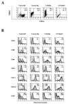
FIG. 1.
FIG. 1.
C. albicans Y form-phagocytosing monocytes do not differentiate into DCs, while C. albicans GT-phagocytosing monocytes differentiate into atypical mature DCs. Human monocytes from a healthy donor were allowed to phagocytose C. albicans Y or GT forms and then induced to differentiate into DCs during a 6-day culture in the presence of GM-CSF and IL-4. DCs derived from noninfected monocytes derived from the same donor and macrophages derived from monocytes cultured for 6 days with M-CSF were used to compare the phenotypes of Y-MoMφs and GT-MoDCs. (A) Dot plots showing the surface expression of CD14 and CD1a of control DCs, control macrophages (control Mφs), Y-MoMφs, and GT-MoDCs. The numbers indicate the percentages of cells in the four quadrants. (B) Histograms of control DCs, control macrophages, Y-MoMφs, and GT-MoDCs that were treated (shaded histograms) or were not treated (unshaded histograms) with LPS. The results obtained with isotypic controls are indicated by dotted lines. The numbers indicate the mean intensities of fluorescence with (positive values) and without (negative values) LPS treatment of cells stained with anti-CD83, anti-CD80, anti-CD86, anti-CD40, or anti-MHC class I and II. For CCR7 staining the percentages of positive cells with (positive values) and without (negative values) LPS treatment are shown. The data for control DCs, Y-MoMφs, and GT-MoDCs are from one experiment that was representative of five independent experiments; the data for control macrophages are representative of the data obtained in two independent experiments.
FIG. 2.
FIG. 2.
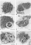
Transmission electron microscopy reveals that monocytes phagocytosing C. albicans Y forms or GTs are induced to differentiate into morphologically distinct cell populations. (a) Control monocyte. (b) Cells cultured for 6 days in the presence of GM-CSF and IL-4, showing the typical DC morphology. (c) Interaction between monocytes and Y forms of C. albicans after incubation for 1 h. The arrow indicates a completely internalized Y cell. (d) Interaction between monocytes and GT forms of C. albicans after incubation for 1 h. The arrows indicate internalized GTs. (e) After 6 days of incubation with the Y forms, numerous digested cells or remnants of fungal cells (arrows) were observed inside monocytes having a macrophage morphology. (f) After 6 days of incubation with the GT forms, the presence of numerous internalized and digested fungal cells (arrows) did not interfere with the morphological differentiation of monocytes into DCs. Bars = 2 μm.
FIG. 3.
FIG. 3.
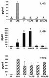
Cells derived from C. albicans-phagocytosing monocytes are unable to produce IL-12p70: IL-12p70, IL-10, and TNF-α secretion in supernatants of cells derived from monocytes infected with Y forms or GTs. Ni, noninfected control DCs; LPS, control DCs stimulated overnight with LPS; Y+LPS and GT+LPS, cells derived from monocytes infected with Y forms and GTs, respectively, which were further stimulated with LPS overnight. An asterisk indicates that the value is significantly different from the value for control DCs stimulated with LPS, as calculated by using analysis of variance and the Student-Newman-Keuls posttest. The results are the means of three independent experiments; the error bars indicate standard deviations.
FIG. 4.
FIG. 4.
Cells derived from Y form- or GT-phagocytosing monocytes are not exhausted and display different patterns of mRNA cytokine expression: reverse transcription-PCR analysis of cells derived from monocytes infected with Y forms or GTs stimulated or not stimulated with LPS in the last 18 h of culture. The data are representative of three experiments. MW, molecular weight.
FIG. 5.
FIG. 5.
Cells derived from C. albicans-phagocytosing monocytes have an impaired antigen presentation function. (A and B) Proliferative response of T lymphocytes from adult peripheral blood (A) or cord blood (B) stimulated with allogeneic DCs (ctr) or with cells derived from monocytes phagocytosing C. albicans Y forms or GTs. Control DCs, Y-MoMφs, and GT-MoDCs were obtained by culturing monocytes from the same healthy donor and were used both with LPS treatment (+LPS) and without LPS treatment in the last 18 h of the 6-day differentiation culture in the presence of GM-CSF and IL-4. The data are from one experiment that was representative of four experiments. (C) Flow cytometric analysis of intracellular cytokine accumulation in CB-Ts after 6 days of coculture with allogeneic APCs. The intracellular cytokine accumulation was detected in CD3-positive cells after 6 days of culture with allogeneic noninfected mature DCs (N.I.) or cells derived from Y form- or GT-infected monocytes. Dot plots show data obtained in experiments in which all APCs were treated overnight with LPS and then extensively washed before incubation with CB-Ts. The numbers in the panels indicate the percentages of cells in the corresponding quadrants. The data are from one experiment that was representative of four experiments.

References

    1. Allavena, P., L. Piemonti, D. Longoni, S. Bernasconi, A. Stoppacciaro, L. Ruco, and A. Mantovani. 1998. IL-10 prevents the differentiation of monocytes to dendritic cells but promotes their maturation to macrophages. Eur. J. Immunol. 28:359-369. - PubMed
    1. Ausiello, C. M., F. Urbani, S. Gessani, G. C. Spagnoli, M. J. Gomez, and A. Cassone. 1993. Cytokine gene expression in human peripheral blood mononuclear cells stimulated by mannoprotein constituents from Candida albicans. Infect. Immun. 61:4105-4111. - PMC - PubMed
    1. Bistoni, F., E. Cenci, A. Mencacci, E. Schiaffella, P. Mosci, P. Puccetti, and L. Romani. 1993. Mucosal and systemic T helper cell function after intragastric colonization of adult mice with Candida albicans. J. Infect. Dis. 168:1449-1457. - PubMed
    1. Cassone, A., F. De Bernardis, C. M. Ausiello, M. J. Gomez, M. Boccanera, R. La Valle, and A. Torosantucci. 1998. Immunogenic and protective Candida albicans constituents. Res. Immunol 149:289-299. (Discussion, 149:504.) - PubMed
    1. Caux, C., C. Massacrier, B. Vanbervliet, B. Dubois, I. Durand, M. Cella, A. Lanzavecchia, and J. Banchereau. 1997. CD34+ hematopoietic progenitors from human cord blood differentiate along two independent dendritic cell pathways in response to granulocyte-macrophage colony-stimulating factor plus tumor necrosis factor alpha. II. Functional analysis. Blood 90:1458-1470. - PubMed

Publication types